- Published on
Clinical Procedures - Exercise Tolerance Testing (ETT)
Indications
• Assessment of chest pain in those with known coronary artery disease (there is no longer a role for ETT in patients presenting with chest pain who do not have a history of coronary artery disease)
• Assessment of haemodynamic response in those with known valvular disease who are asymptomatic
• Diagnosis of exertionally induced arrhythmias or syncope.
Contraindications
• Any undiagnosed or previously unknown murmur (patient should undergo echocardiogram first)
• Severe aortic stenosis (risk of syncope)
• Hypertrophic cardiomyopathy with significant outflow obstruction (risk of syncope)
• Severe hyper- or hypotension
• Unstable angina (should undergo coronary angiography)
• Known severe left main stem disease
• Untreated congestive cardiac failure
• Complete heart block
• Aortic aneurysm
• Acute myocarditis or pericarditis
• Any recent pyrexial or 'flu-like' illness.
Procedure
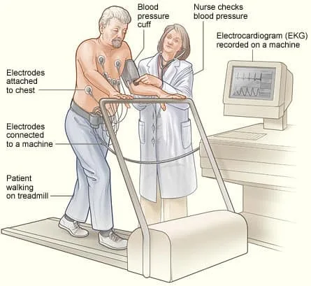
• ECG electrodes are put on the patient's chest and a sphygmomanometer cuff on an arm
• The patient is asked to walk on a treadmill connected to the computer whilst their ECG, blood pressure, and heart rate are monitored. The speed and incline of the treadmill increase according to set protocols:
• Bruce protocol: for assessment of physically fit and stable patients with suspected coronary artery disease, 7 stages starting at a 10% gradient at 1.7mph and increasing to 22% gradient and 6mph
• Modified Bruce protocol: used in elderly patients or those who have been stabilized after a suspected episode of unstable angina.
Starts at 1.7mph and 0% gradient and increases the gradient slowly to 10%
• Termination of the test depends on the results seen (see box).
Risks
• Risks are those associated with exercise and include:
• Arrhythmia, cardiac ischaemia, myocardial infarction, syncope.
Patient Preparation
• No specific preparation is required. Patients are asked not to eat or drink for 3 hours prior to the test
• Comfortable clothing and shoes should be worn.
Indications for termination of procedure
• Patient requests to stop
• Symptoms: fatigue, angina, dizziness, significant breathlessness
• Signs: drop in oxygen saturations <94%, target heart rate achieved, hypotension during exercise (e.g. BP <100mmg), significant hypertension (e.g. BP >200mmHg)
• ECG: any atrial or ventricular arrhythmia, frequent ventricular ectopics, new AV or bundle branch block, ST segment shift >1mm.
Causes of false positive results or low specificity
• Often due to difficulty interpreting results as result of resting ST segment abnormalities:
•Wolf-Parkinson-White syndrome, left bundle branch block, atrial fibrillation, left ventricular hypertrophy, digoxin therapy, hyperventilation, biochemical electrolyte abnormalities (e.g. hypo- or hyperkalaemia), cardiomyopathies, left ventricular outflow obstruction
Beta-blocker therapy prevents the appropriate heart rate/blood
pressure response during testing.
Indications
• Assessment of chest pain in those with known coronary artery disease (there is no longer a role for ETT in patients presenting with chest pain who do not have a history of coronary artery disease)
• Assessment of haemodynamic response in those with known valvular disease who are asymptomatic
• Diagnosis of exertionally induced arrhythmias or syncope.
Contraindications
• Any undiagnosed or previously unknown murmur (patient should undergo echocardiogram first)
• Severe aortic stenosis (risk of syncope)
• Hypertrophic cardiomyopathy with significant outflow obstruction (risk of syncope)
• Severe hyper- or hypotension
• Unstable angina (should undergo coronary angiography)
• Known severe left main stem disease
• Untreated congestive cardiac failure
• Complete heart block
• Aortic aneurysm
• Acute myocarditis or pericarditis
• Any recent pyrexial or 'flu-like' illness.
Procedure
• ECG electrodes are put on the patient's chest and a sphygmomanometer cuff on an arm
• The patient is asked to walk on a treadmill connected to the computer whilst their ECG, blood pressure, and heart rate are monitored. The speed and incline of the treadmill increase according to set protocols:
• Bruce protocol: for assessment of physically fit and stable patients with suspected coronary artery disease, 7 stages starting at a 10% gradient at 1.7mph and increasing to 22% gradient and 6mph
• Modified Bruce protocol: used in elderly patients or those who have been stabilized after a suspected episode of unstable angina.
Starts at 1.7mph and 0% gradient and increases the gradient slowly to 10%
• Termination of the test depends on the results seen (see box).
Risks
• Risks are those associated with exercise and include:
• Arrhythmia, cardiac ischaemia, myocardial infarction, syncope.
Patient Preparation
• No specific preparation is required. Patients are asked not to eat or drink for 3 hours prior to the test
• Comfortable clothing and shoes should be worn.
Indications for termination of procedure
• Patient requests to stop
• Symptoms: fatigue, angina, dizziness, significant breathlessness
• Signs: drop in oxygen saturations <94%, target heart rate achieved, hypotension during exercise (e.g. BP <100mmg), significant hypertension (e.g. BP >200mmHg)
• ECG: any atrial or ventricular arrhythmia, frequent ventricular ectopics, new AV or bundle branch block, ST segment shift >1mm.
Causes of false positive results or low specificity
• Often due to difficulty interpreting results as result of resting ST segment abnormalities:
•Wolf-Parkinson-White syndrome, left bundle branch block, atrial fibrillation, left ventricular hypertrophy, digoxin therapy, hyperventilation, biochemical electrolyte abnormalities (e.g. hypo- or hyperkalaemia), cardiomyopathies, left ventricular outflow obstruction
Beta-blocker therapy prevents the appropriate heart rate/blood
pressure response during testing.
0 Comments
