- Published on
Clinical Procedures - Incision and Drainage
Equipment
• Mask, eye protection, and gloves
• Antiseptic solution and drapes
• Scalpel
• Microbiology swab
• Forceps
• Curette
• 2 x kidney dish (1 dirty, 1 for wash solution)
• 50mL syringe
• Swabs
• Normal saline
• Ribbon pack e.g. Aquacel®.
Procedure
• Introduce yourself, confirm the patient's identity, explain the procedure, and obtain informed written consent
• Position patient to allow easy access to abscess area
• Wear mask and protective glasses as the pus can be under pressure and spray!
• Clean hands with surgical scrub method and put on sterile gloves
• Clean and drape area appropriately
• Hold kidney dish below abscess to collect pus
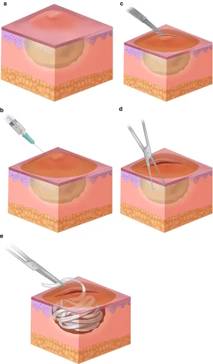
• Make an incision with the scalpel over 'pointed' area or most fluctuant point of abscess:
• Try to incise along natural skin lines. This can be difficult when there is distortion of tissue due to abscess swelling
• Take microbiology swab of pus
• Extend incision to allow good drainage of the cavity
• Insert a gloved finger and gently break down any loculations
• Perform a curettage of the wound to remove the thick abscess capsule if present
• Excise any necrotic skin using forceps and scalpel
• Wash the cavity with normal saline until wash runs clear.
The volume needed depends on the size of the abscess
• Pack wound loosely with ribbon pack and apply dressing
• Clean area around the wound
• If the patient is systemically unwell, immunocompromised, or if cellulitis is spreading from the abscess area, a course of IV antibiotics will need to be given.
Follow-up
• The pack should be changed at 24 hours and the wound inspected
• If sending the patient home, ask district nurses or practice nurse to assess and change pack.
Documentation
• Date, time, indication, informed consent obtained
• Site and size of abscess, cellulitis, and observations
• Anaesthetic technique (e.g. local or general)
• Incision technique
• Findings (dimensions of cavity, loculations, amount of pus)
• Microbiology swab taken?
• Haemostasis achieved?
• Volume of wash solution used
• Type of pack used
• Follow-up instructions and antibiotics (if needed)
• Advice given to patient
• Signature, printed name, and contact details.
Indications
• Painful swollen area
• Spreading cellulitis
• Systemically unwell
• Failure of antibiotic treatment.
Contraindications
• Pulsatile swelling (e.g. false or true aneurysm).
Risks
• Spread or recurrence of infection, bleeding
• Scar
• Further surgery.
Procedures Tips
Necrotizing fasciitis/Fournier's gangrene
• Greatest risk in immunocompromised and peri-anal or scrotal wounds
• Possible signs include:
• Systemically unwell
• Rapidly spreading erythema ‡ skin necrosis
• Surgical emphysema
• Easy passage of finger between skin and fascia planes
• Yellowish-green necrotic fascia.
Perianal abscesses
• Perform a digital rectal examination feeling for a 'grain of rice' in the anal canal suggesting a fistula opening or bulge from a collection of pus
• Perform a rigid sigmoidoscopy looking for pus or rectal tumours
• If a fistula is suspected, probing for a tract may create false tracts
• Follow-up microbiology:
• If skin bacteria (e.g. S. aureus or S. epidermidis), the abscess is probably secondary to an infected skin follicle
• If gut bacteria, (e.g. E. coli), the abscess is probably due to an infected peri-anal gland. Increased chance of fistula formation and follow-up to check wound healing ‡ referral to colorectal specialist is advised.
Groin abscesses
• Suspect false aneurysm if the patient has been attempting IV injections at the site
• Check for pulsatility and request ultrasound to investigate further.
Equipment
• Mask, eye protection, and gloves
• Antiseptic solution and drapes
• Scalpel
• Microbiology swab
• Forceps
• Curette
• 2 x kidney dish (1 dirty, 1 for wash solution)
• 50mL syringe
• Swabs
• Normal saline
• Ribbon pack e.g. Aquacel®.
Procedure
• Introduce yourself, confirm the patient's identity, explain the procedure, and obtain informed written consent
• Position patient to allow easy access to abscess area
• Wear mask and protective glasses as the pus can be under pressure and spray!
• Clean hands with surgical scrub method and put on sterile gloves
• Clean and drape area appropriately
• Hold kidney dish below abscess to collect pus
• Make an incision with the scalpel over 'pointed' area or most fluctuant point of abscess:
• Try to incise along natural skin lines. This can be difficult when there is distortion of tissue due to abscess swelling
• Take microbiology swab of pus
• Extend incision to allow good drainage of the cavity
• Insert a gloved finger and gently break down any loculations
• Perform a curettage of the wound to remove the thick abscess capsule if present
• Excise any necrotic skin using forceps and scalpel
• Wash the cavity with normal saline until wash runs clear.
The volume needed depends on the size of the abscess
• Pack wound loosely with ribbon pack and apply dressing
• Clean area around the wound
• If the patient is systemically unwell, immunocompromised, or if cellulitis is spreading from the abscess area, a course of IV antibiotics will need to be given.
Follow-up
• The pack should be changed at 24 hours and the wound inspected
• If sending the patient home, ask district nurses or practice nurse to assess and change pack.
Documentation
• Date, time, indication, informed consent obtained
• Site and size of abscess, cellulitis, and observations
• Anaesthetic technique (e.g. local or general)
• Incision technique
• Findings (dimensions of cavity, loculations, amount of pus)
• Microbiology swab taken?
• Haemostasis achieved?
• Volume of wash solution used
• Type of pack used
• Follow-up instructions and antibiotics (if needed)
• Advice given to patient
• Signature, printed name, and contact details.
Indications
• Painful swollen area
• Spreading cellulitis
• Systemically unwell
• Failure of antibiotic treatment.
Contraindications
• Pulsatile swelling (e.g. false or true aneurysm).
Risks
• Spread or recurrence of infection, bleeding
• Scar
• Further surgery.
Procedures Tips
Necrotizing fasciitis/Fournier's gangrene
• Greatest risk in immunocompromised and peri-anal or scrotal wounds
• Possible signs include:
• Systemically unwell
• Rapidly spreading erythema ‡ skin necrosis
• Surgical emphysema
• Easy passage of finger between skin and fascia planes
• Yellowish-green necrotic fascia.
Perianal abscesses
• Perform a digital rectal examination feeling for a 'grain of rice' in the anal canal suggesting a fistula opening or bulge from a collection of pus
• Perform a rigid sigmoidoscopy looking for pus or rectal tumours
• If a fistula is suspected, probing for a tract may create false tracts
• Follow-up microbiology:
• If skin bacteria (e.g. S. aureus or S. epidermidis), the abscess is probably secondary to an infected skin follicle
• If gut bacteria, (e.g. E. coli), the abscess is probably due to an infected peri-anal gland. Increased chance of fistula formation and follow-up to check wound healing ‡ referral to colorectal specialist is advised.
Groin abscesses
• Suspect false aneurysm if the patient has been attempting IV injections at the site
• Check for pulsatility and request ultrasound to investigate further.
0 Comments
