- Published on
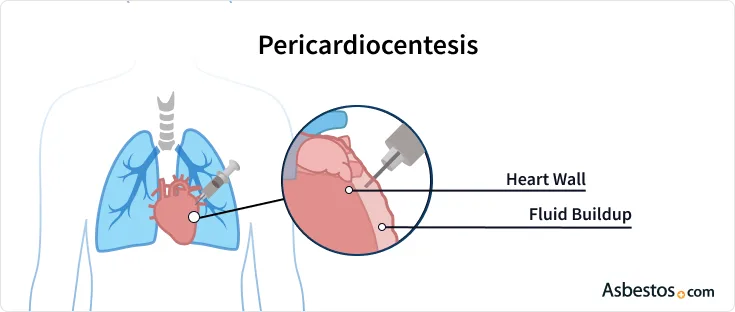
Clinical Procedures – Pericardiocentesis
Equipment
• Echocardiogram machine and sterile probe cover
• Pericardial drain kit (14 gauge needle, syringe, guidewire, pigtail catheter and drain)
• Sterile drape and towels
• lodine solution
• Sterile gloves and gown
• Local anaesthetic (1% lidocaine)
• 2 x 10mL syringe
• Orange/blue/green needles
• Sterile gauze
• 50mL syringe
• 3-way tap
• Suture, scissors, sticky dressing (e.g. Tegaderm®).
You will also need:
• IV access
• ECG monitoring
• Access to 'crash' trolley (defibrillator and emergency drugs).
Procedure
• Introduce yourself, explain the procedure, and obtain informed written consent
• Ensure IV access, ECG monitoring, normal clotting, and access to resuscitation equipment
• (Consider light sedation)
• Position patient supine with 20-30° head tilt
• Ensure all equipment is sterile and laid out on sterile trolley
• Wash hands using surgical scrub technique and put on the sterile gown and gloves
• Clean and drape site at the inferior border of the sternum:
• The point of needle insertion is immediately below and to the left of the xiphisternum
• Confirm location of effusion using echocardiogram machine with sterile probe cover
• Infiltrate overlying skin and subcutaneous tissue with 1% lidocaine. (Always aspirate before each injection.)
• Attach the 10mL syringe attached to the 14G needle
• Insert the needle between the xiphisternum and left costal margin advancing slowly at 35° to the patient and aiming towards the patient's left shoulder. Aspirate continuously as the needle advances:
• Pericardial fluid is usually aspirated at about 6 8cm depth
Depending on the size of the pericardial effusion and indication for the procedure, you may wish to attach the 50mL syringe and aspirate fluid to send for diagnostic purposes
• A modified Seldinger technique should now be used to insert the pericardial drain
• Once pericardial fluid is aspirated, hold the needle in position, remove the syringe, and insert the guidewire slowly through the needle into the pericardial space
• Remove the needle, holding the wire in place at all times
• Pass the catheter over the wire into the pericardial space
• Once the catheter position is confirmed on echo, remove the wire and attach the 3-way tap and drain bag
• Suture the drain in place and dress to maintain sterility
• Request a CXR to exclude iatrogenic pneumothorax.
Documentation
• Date, time, and place
• Name and grade of person who performed the procedure (and anyone who supervised)
• Consent obtained (enclose copy of consent form)
• Aseptic technique used and volume of anesthetic used
• Approach taken and anatomy confirmed by echocardiogram
• Any difficulties, i.e. "first pass' or 'second attempt' etc.
• Appearance of pericardial fluid aspirated
• Volume of pericardial fluid aspirated.
Indications
• Treatment of cardiac tamponade
• Symptomatic pericardial effusion
• Diagnostic pericardiocentesis
• During CPR to exclude cardiac tamponade as a reversible cause of cardiac arrest.
Contraindications
• Cardiac tamponade secondary to cardiac trauma or aortic dissection (surgical intervention is preferable)
• Recurrent pericardial effusions (surgical pericardial window indicated).
Risks
• Pneumothorax
• Myocardial perforation
• Cardiac tamponade
• Coronary artery laceration
• Cardiac arrhythmias
• Intra-abdominal trauma (especially to liver)
• Haemorrhage
• Infection
• Acute pulmonary oedema
• Failure of procedure
• Death.
Procedure Tips
• Pericardiocentesis should be performed by a trained doctor (either cardiologist or thoracic surgeon usually) preferably in a sterile environment (theatre or the cardiac catheterization lab) and under echocardiographic guidance, with access to full resuscitation equipment
• The only exception is during cardiopulmonary resuscitation when pericardiocentesis is performed as an emergency to exclude cardiac tamponade as a reversible cause of cardiac arrest
• Always check the patient's clotting before commencing pericardiocentesis
• The clinician who performed the procedure should confirm the position of the drain using echo
• Always request a post-procedure chest radiograph to exclude iatrogenic pneumothorax.
Equipment
• Echocardiogram machine and sterile probe cover
• Pericardial drain kit (14 gauge needle, syringe, guidewire, pigtail catheter and drain)
• Sterile drape and towels
• lodine solution
• Sterile gloves and gown
• Local anaesthetic (1% lidocaine)
• 2 x 10mL syringe
• Orange/blue/green needles
• Sterile gauze
• 50mL syringe
• 3-way tap
• Suture, scissors, sticky dressing (e.g. Tegaderm®).
You will also need:
• IV access
• ECG monitoring
• Access to 'crash' trolley (defibrillator and emergency drugs).
Procedure
• Introduce yourself, explain the procedure, and obtain informed written consent
• Ensure IV access, ECG monitoring, normal clotting, and access to resuscitation equipment
• (Consider light sedation)
• Position patient supine with 20-30° head tilt
• Ensure all equipment is sterile and laid out on sterile trolley
• Wash hands using surgical scrub technique and put on the sterile gown and gloves
• Clean and drape site at the inferior border of the sternum:
• The point of needle insertion is immediately below and to the left of the xiphisternum
• Confirm location of effusion using echocardiogram machine with sterile probe cover
• Infiltrate overlying skin and subcutaneous tissue with 1% lidocaine. (Always aspirate before each injection.)
• Attach the 10mL syringe attached to the 14G needle
• Insert the needle between the xiphisternum and left costal margin advancing slowly at 35° to the patient and aiming towards the patient's left shoulder. Aspirate continuously as the needle advances:
• Pericardial fluid is usually aspirated at about 6 8cm depth
Depending on the size of the pericardial effusion and indication for the procedure, you may wish to attach the 50mL syringe and aspirate fluid to send for diagnostic purposes
• A modified Seldinger technique should now be used to insert the pericardial drain
• Once pericardial fluid is aspirated, hold the needle in position, remove the syringe, and insert the guidewire slowly through the needle into the pericardial space
• Remove the needle, holding the wire in place at all times
• Pass the catheter over the wire into the pericardial space
• Once the catheter position is confirmed on echo, remove the wire and attach the 3-way tap and drain bag
• Suture the drain in place and dress to maintain sterility
• Request a CXR to exclude iatrogenic pneumothorax.
Documentation
• Date, time, and place
• Name and grade of person who performed the procedure (and anyone who supervised)
• Consent obtained (enclose copy of consent form)
• Aseptic technique used and volume of anesthetic used
• Approach taken and anatomy confirmed by echocardiogram
• Any difficulties, i.e. "first pass' or 'second attempt' etc.
• Appearance of pericardial fluid aspirated
• Volume of pericardial fluid aspirated.
Indications
• Treatment of cardiac tamponade
• Symptomatic pericardial effusion
• Diagnostic pericardiocentesis
• During CPR to exclude cardiac tamponade as a reversible cause of cardiac arrest.
Contraindications
• Cardiac tamponade secondary to cardiac trauma or aortic dissection (surgical intervention is preferable)
• Recurrent pericardial effusions (surgical pericardial window indicated).
Risks
• Pneumothorax
• Myocardial perforation
• Cardiac tamponade
• Coronary artery laceration
• Cardiac arrhythmias
• Intra-abdominal trauma (especially to liver)
• Haemorrhage
• Infection
• Acute pulmonary oedema
• Failure of procedure
• Death.
Procedure Tips
• Pericardiocentesis should be performed by a trained doctor (either cardiologist or thoracic surgeon usually) preferably in a sterile environment (theatre or the cardiac catheterization lab) and under echocardiographic guidance, with access to full resuscitation equipment
• The only exception is during cardiopulmonary resuscitation when pericardiocentesis is performed as an emergency to exclude cardiac tamponade as a reversible cause of cardiac arrest
• Always check the patient's clotting before commencing pericardiocentesis
• The clinician who performed the procedure should confirm the position of the drain using echo
• Always request a post-procedure chest radiograph to exclude iatrogenic pneumothorax.
0 Comments
