- Published on
Kembara Xtra - Medicine - Atypical Mole Syndrome
BASICS
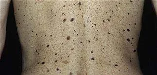
Atypical mole syndrome (AMS), also referred to as dysplastic nevi syndrome (DNS), B-K mole syndrome, Clark nevi syndrome, or familial atypical multiple mole melanoma (FAMMM) syndrome, is a condition that is characterized by an abundance of pigmented nevi with architectural disorder. These nevi can develop sporadically or through inheritance and are linked to an increased risk of developing melanoma.
DESCRIPTION There is no agreed-upon set of standards for AMS.
The number of clinically unusual nevi included in the elevated total body nevi count is often >50 and frequently >100.
- More prevalent in hereditary AMS than sporadic atypical nevi, which might be as low as 10.
Melanoma risk increased - Up to 90% of cases by age 80 in some high-risk individuals The median age of diagnosis for melanoma in AMS is 10 to 20 years earlier than the general population, with documented cases of melanoma as early as in the second and third decades of life. More often than not, these tumors arise de novo rather than from an existing nevus.
EPIDEMIOLOGY
Incidence
Due to phenotypic heterogeneity and a lack of evidence, uncertain
Prevalence
2% to 8% of fair-skinned adults and people who are exposed to a lot of UV radiation are affected by this condition.
PATHOPHYSIOLOGY AND ETIOLOGY
Multiple melanomas and familial DNS have both been linked to mutations in the CDKN2A protein. The CDKN2A gene on 9p21 codes for the proteins p16 and p14. P16 binds to CDK4/6 and inhibits the CDKcyclin D interaction required for cell cycle progression from G1 to S. P14 has a similar function to p16 by stabilizing the tumor suppressor protein p53 in the G1 phase of the cell cycle.
There are no discernible somatic mutation patterns in sporadic occurrences. Familial cases of germline CDKN2A mutations are transmitted in an autosomal dominant manner.
Genetically speaking, autosomal dominant inheritance is seen in 25–40% of hereditary cases, with varying expressivity and partial penetrance.
RISK ELEMENTS
Multiple nevi or a family history of melanoma, sun exposure, neonatal blue-light phototherapy, and a history of severe sunburns
GENERAL PREVENTION Primary prevention with sun avoidance and sun protection Secondary prevention of melanoma with routine skin checks, biopsy of questionable lesions, and risk reduction for the environment as stated above
COMMONLY ASSOCIATED CONDITIONS Pancreatic cancer in CDKN2A mutation Malignant melanoma, including ocular melanoma Ocular nevi
DIAGNOSIS
There have been several proposed classification schemes for the clinical diagnosis of AMS. Although not widely accepted, the NIH's diagnostic criteria call for the three characteristics of (i) malignant melanoma in 1 first- or second-degree relatives; (ii) numerous melanocytic nevi (often >50); and (iii) nevi with specific histologic characteristics.
HISTORY
Large quantity of nevi; Congenital nevi; Changing lesions: bleeding, scale, size, texture, nonhealing; Hyper- or Hypopigmentation
Exposure to the sun, earlier skin biopsies, and earlier melanoma
Immunosuppression (such as pancreatic cancer, AIDS, or chemotherapy); First- or second-degree relatives with:
- Pancreatic cancer - AMS - Melanoma
Full-body skin examinations with photographs to monitor developing nevi
Aiming to distinguish melanoma from an atypical mole (AM), the ABCDE acronym stands for the following skin lesions: Asymmetry, irregular borders, color variation, a diameter of at least 6 mm, and an evolving lesion
- AM is frequently outlined as having at least two additional characteristics and a diameter of less than 5 mm.
– When a lesion's diameter is greater than 6 mm, the specificity for melanoma is enhanced. "Ugly duckling sign" (2)[B]:
- Melanoma screening technique to detect malignant nevi deviating from the prevalent nevus pattern when a large number of atypical nevi are present, improving the accuracy of melanoma diagnosis The following three [C] are the most typical dermoscopy findings for AM:
The most frequent reticular pattern is uniform pigmentation, which is followed by multifocal hypo/hyperpigmentation.
uniform brown globules
- Pigmentation with abrupt end and central heterogeneity The following cutaneous characteristics are more indicative of melanoma:
- Discolorations
Whitish veil, homogenous patches dispersed unevenly, in many locations, or >25% of the overall lesion, and less than four colors
DIFFERENTIAL DIAGNOSIS Melanoma, Seborrheic Keratosis, Dermatofibroma, Lentigo, Pigmented Actinic Keratosis, Pigmented Basal Cell Carcinoma, and Blue Rubber Bleb Nevus Syndrome are among the diagnoses that can be made.
DETECTION & INTERPRETATION OF DIAGNOSIS
Histopathology and biopsy results are used to confirm the diagnosis when the history and physical examination raise suspicions.
Initial examinations (lab, imaging)
Dermoscopy can be performed for a more thorough examination of nevi to help differentiate between benign and malignant lesions and to further categorize to any of the 11 subtypes; however, the degree of success is reliant on the examiner's skill.
When separating AM from melanoma, reflectance confocal microscopy (RCM) may be more accurate than dermoscopy.
Total body photography may help in the evaluation of developing nevi as well as in documenting new nevi when the total number of nevi is high and following individual nevus is problematic.
Tests in the Future & Special Considerations
For CDKN2A mutations, genetic testing is accessible, but it is not advised outside of research investigations because the results cannot be effectively used for management or surveillance.
Other/Diagnostic Procedures
Any lesion where melanoma cannot be ruled out requires a biopsy.
In order to properly measure the depth of the lesion, a biopsy must be performed on the entire lesion down to the fat layer, leaving just a small 1- to 3-mm margin of healthy skin.
- Punch or elliptical excisional biopsy offers the most precise diagnosis and ought to be carried out whenever practical.
- Scoop shave biopsy is another option, however it must be done carefully to avoid transecting the lesion.
Reexcision of mild to moderately dysplastic nevi with positive margins may not alter the pathologic diagnosis or prognosis (studies inconclusive) (6)[A], but for severely dysplastic nevi, reexcision with surgical margins of 2 to 5 mm may be considered.
Interpretation of Tests
The phrase "dysplastic nevus" is better used to describe a histologic diagnosis. The presence of rete ridge fusion, dermal fibrosis, neovascularization, interstitial lymphocytic inflammation, and melanocyte proliferation in the dermoepidermal interface that extends through at least three rete ridges in a particular pattern are possible characteristics.
TREATMENT /MEDICATION
There is no evidence that any drugs can treat AMS.
QUESTIONS FOR REFERENCE
For individuals who are at high risk for melanoma, a dermatologist should perform routine skin exams. individuals with papilledema, ocular nevi, or both should have ophthalmologic examinations. If there is a significant family history of pancreatic cancer, a genetics study group should be consulted.
ADVANCED THERAPIES
The ineffective use of topical chemo- and immunotherapies to treat AMs.
Because laser therapy for melanocytic nevi is risky and unsuccessful, it should be avoided.
SURGICAL AND OTHER PROCEDURE
The majority of melanomas in AMS emerge de novo on healthy skin; consequently, surgical removal of all atypical nevi is not advised as it has little therapeutic utility and is not financially advantageous. Additionally, excision of all atypical nevi produces subpar cosmetic results and gives the patient a false sense of security.
Biopsies or surgical removal should be performed on lesions thought to be melanoma.
CONTINUING CARE AFTERCARE RECOMMENDATIONS
Maintain in contact with a dermatologist or other medical professional with experience evaluating atypical nevi:
Starting at adolescence, a full body skin examination should be performed every six months. Once nevi are stable, it may be lowered to once a year.
Total body photography to monitor the development of new nevi and their progression over time.
Ocular examination for patients with familial AMS, dermoscopic evaluation of worrisome lesions, excision of suspicious lesions
Patient Monitoring Regular skin examinations
EDUCATION OF PATIENTS
Encourage young adults with fair skin to limit their exposure to UV radiation to lower their chance of developing skin cancer (USPSTF grade B).
Fair skin: light-colored hair, eyes, or skin, freckles
Inform people about how to avoid the sun, apply sunscreen correctly, use protective clothes (such hats), avoid tanning booths, and prevent sunburns.
To evaluate nevi and spot probable melanomas, teach the "ABCDE" mnemonic and the "ugly duckling sign".
Give instructions on how to conduct a skin self-exam.
PROGNOSIS
Most AM either stagnate or regress.
● Over the years, numerous categorization systems have been created to identify the risk of melanoma in individuals with AMS. The riskiest group of people are those with a family history of melanoma. It is possible to use the Rigel categorization system in a clinical environment. In accordance with the incidence of melanoma, points are stratified as follows: 1 point is awarded for a personal history of melanoma; 2 points are awarded for each family member who has the disease (defined as a modified nuclear family made up of first-degree relatives plus grandparents and uncles/aunts):
Score = 1, Rigel group 1, 10% risk; Score = 2, Rigel group 2, 15% risk; Score 3, Rigel group 3, 50% risk; Score = 0, Rigel group 0, 6% 25-year accumulated risk for melanoma
Additionally, the CDKN2A mutation has been linked to a 60–90% risk of melanoma by the age of 80 and a 17% risk of pancreatic cancer by the age of 75.
Malignant melanoma and subpar cosmetic results from biopsies are complications.
BASICS
Atypical mole syndrome (AMS), also referred to as dysplastic nevi syndrome (DNS), B-K mole syndrome, Clark nevi syndrome, or familial atypical multiple mole melanoma (FAMMM) syndrome, is a condition that is characterized by an abundance of pigmented nevi with architectural disorder. These nevi can develop sporadically or through inheritance and are linked to an increased risk of developing melanoma.
DESCRIPTION There is no agreed-upon set of standards for AMS.
The number of clinically unusual nevi included in the elevated total body nevi count is often >50 and frequently >100.
- More prevalent in hereditary AMS than sporadic atypical nevi, which might be as low as 10.
Melanoma risk increased - Up to 90% of cases by age 80 in some high-risk individuals The median age of diagnosis for melanoma in AMS is 10 to 20 years earlier than the general population, with documented cases of melanoma as early as in the second and third decades of life. More often than not, these tumors arise de novo rather than from an existing nevus.
EPIDEMIOLOGY
Incidence
Due to phenotypic heterogeneity and a lack of evidence, uncertain
Prevalence
2% to 8% of fair-skinned adults and people who are exposed to a lot of UV radiation are affected by this condition.
PATHOPHYSIOLOGY AND ETIOLOGY
Multiple melanomas and familial DNS have both been linked to mutations in the CDKN2A protein. The CDKN2A gene on 9p21 codes for the proteins p16 and p14. P16 binds to CDK4/6 and inhibits the CDKcyclin D interaction required for cell cycle progression from G1 to S. P14 has a similar function to p16 by stabilizing the tumor suppressor protein p53 in the G1 phase of the cell cycle.
There are no discernible somatic mutation patterns in sporadic occurrences. Familial cases of germline CDKN2A mutations are transmitted in an autosomal dominant manner.
Genetically speaking, autosomal dominant inheritance is seen in 25–40% of hereditary cases, with varying expressivity and partial penetrance.
RISK ELEMENTS
Multiple nevi or a family history of melanoma, sun exposure, neonatal blue-light phototherapy, and a history of severe sunburns
GENERAL PREVENTION Primary prevention with sun avoidance and sun protection Secondary prevention of melanoma with routine skin checks, biopsy of questionable lesions, and risk reduction for the environment as stated above
COMMONLY ASSOCIATED CONDITIONS Pancreatic cancer in CDKN2A mutation Malignant melanoma, including ocular melanoma Ocular nevi
DIAGNOSIS
There have been several proposed classification schemes for the clinical diagnosis of AMS. Although not widely accepted, the NIH's diagnostic criteria call for the three characteristics of (i) malignant melanoma in 1 first- or second-degree relatives; (ii) numerous melanocytic nevi (often >50); and (iii) nevi with specific histologic characteristics.
HISTORY
Large quantity of nevi; Congenital nevi; Changing lesions: bleeding, scale, size, texture, nonhealing; Hyper- or Hypopigmentation
Exposure to the sun, earlier skin biopsies, and earlier melanoma
Immunosuppression (such as pancreatic cancer, AIDS, or chemotherapy); First- or second-degree relatives with:
- Pancreatic cancer - AMS - Melanoma
Full-body skin examinations with photographs to monitor developing nevi
Aiming to distinguish melanoma from an atypical mole (AM), the ABCDE acronym stands for the following skin lesions: Asymmetry, irregular borders, color variation, a diameter of at least 6 mm, and an evolving lesion
- AM is frequently outlined as having at least two additional characteristics and a diameter of less than 5 mm.
– When a lesion's diameter is greater than 6 mm, the specificity for melanoma is enhanced. "Ugly duckling sign" (2)[B]:
- Melanoma screening technique to detect malignant nevi deviating from the prevalent nevus pattern when a large number of atypical nevi are present, improving the accuracy of melanoma diagnosis The following three [C] are the most typical dermoscopy findings for AM:
The most frequent reticular pattern is uniform pigmentation, which is followed by multifocal hypo/hyperpigmentation.
uniform brown globules
- Pigmentation with abrupt end and central heterogeneity The following cutaneous characteristics are more indicative of melanoma:
- Discolorations
Whitish veil, homogenous patches dispersed unevenly, in many locations, or >25% of the overall lesion, and less than four colors
DIFFERENTIAL DIAGNOSIS Melanoma, Seborrheic Keratosis, Dermatofibroma, Lentigo, Pigmented Actinic Keratosis, Pigmented Basal Cell Carcinoma, and Blue Rubber Bleb Nevus Syndrome are among the diagnoses that can be made.
DETECTION & INTERPRETATION OF DIAGNOSIS
Histopathology and biopsy results are used to confirm the diagnosis when the history and physical examination raise suspicions.
Initial examinations (lab, imaging)
Dermoscopy can be performed for a more thorough examination of nevi to help differentiate between benign and malignant lesions and to further categorize to any of the 11 subtypes; however, the degree of success is reliant on the examiner's skill.
When separating AM from melanoma, reflectance confocal microscopy (RCM) may be more accurate than dermoscopy.
Total body photography may help in the evaluation of developing nevi as well as in documenting new nevi when the total number of nevi is high and following individual nevus is problematic.
Tests in the Future & Special Considerations
For CDKN2A mutations, genetic testing is accessible, but it is not advised outside of research investigations because the results cannot be effectively used for management or surveillance.
Other/Diagnostic Procedures
Any lesion where melanoma cannot be ruled out requires a biopsy.
In order to properly measure the depth of the lesion, a biopsy must be performed on the entire lesion down to the fat layer, leaving just a small 1- to 3-mm margin of healthy skin.
- Punch or elliptical excisional biopsy offers the most precise diagnosis and ought to be carried out whenever practical.
- Scoop shave biopsy is another option, however it must be done carefully to avoid transecting the lesion.
Reexcision of mild to moderately dysplastic nevi with positive margins may not alter the pathologic diagnosis or prognosis (studies inconclusive) (6)[A], but for severely dysplastic nevi, reexcision with surgical margins of 2 to 5 mm may be considered.
Interpretation of Tests
The phrase "dysplastic nevus" is better used to describe a histologic diagnosis. The presence of rete ridge fusion, dermal fibrosis, neovascularization, interstitial lymphocytic inflammation, and melanocyte proliferation in the dermoepidermal interface that extends through at least three rete ridges in a particular pattern are possible characteristics.
TREATMENT /MEDICATION
There is no evidence that any drugs can treat AMS.
QUESTIONS FOR REFERENCE
For individuals who are at high risk for melanoma, a dermatologist should perform routine skin exams. individuals with papilledema, ocular nevi, or both should have ophthalmologic examinations. If there is a significant family history of pancreatic cancer, a genetics study group should be consulted.
ADVANCED THERAPIES
The ineffective use of topical chemo- and immunotherapies to treat AMs.
Because laser therapy for melanocytic nevi is risky and unsuccessful, it should be avoided.
SURGICAL AND OTHER PROCEDURE
The majority of melanomas in AMS emerge de novo on healthy skin; consequently, surgical removal of all atypical nevi is not advised as it has little therapeutic utility and is not financially advantageous. Additionally, excision of all atypical nevi produces subpar cosmetic results and gives the patient a false sense of security.
Biopsies or surgical removal should be performed on lesions thought to be melanoma.
CONTINUING CARE AFTERCARE RECOMMENDATIONS
Maintain in contact with a dermatologist or other medical professional with experience evaluating atypical nevi:
Starting at adolescence, a full body skin examination should be performed every six months. Once nevi are stable, it may be lowered to once a year.
Total body photography to monitor the development of new nevi and their progression over time.
Ocular examination for patients with familial AMS, dermoscopic evaluation of worrisome lesions, excision of suspicious lesions
Patient Monitoring Regular skin examinations
EDUCATION OF PATIENTS
Encourage young adults with fair skin to limit their exposure to UV radiation to lower their chance of developing skin cancer (USPSTF grade B).
Fair skin: light-colored hair, eyes, or skin, freckles
Inform people about how to avoid the sun, apply sunscreen correctly, use protective clothes (such hats), avoid tanning booths, and prevent sunburns.
To evaluate nevi and spot probable melanomas, teach the "ABCDE" mnemonic and the "ugly duckling sign".
Give instructions on how to conduct a skin self-exam.
PROGNOSIS
Most AM either stagnate or regress.
● Over the years, numerous categorization systems have been created to identify the risk of melanoma in individuals with AMS. The riskiest group of people are those with a family history of melanoma. It is possible to use the Rigel categorization system in a clinical environment. In accordance with the incidence of melanoma, points are stratified as follows: 1 point is awarded for a personal history of melanoma; 2 points are awarded for each family member who has the disease (defined as a modified nuclear family made up of first-degree relatives plus grandparents and uncles/aunts):
Score = 1, Rigel group 1, 10% risk; Score = 2, Rigel group 2, 15% risk; Score 3, Rigel group 3, 50% risk; Score = 0, Rigel group 0, 6% 25-year accumulated risk for melanoma
Additionally, the CDKN2A mutation has been linked to a 60–90% risk of melanoma by the age of 80 and a 17% risk of pancreatic cancer by the age of 75.
Malignant melanoma and subpar cosmetic results from biopsies are complications.

0 Comments
