- Published on
Kembara Xtra - Medicine - Erythema Nodosum
Introduction
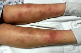
Multiple, bilateral, erythematous, tender nodules in a typically pretibial distribution that undergo a characteristic pattern of color changes, similar to that seen in bruises. A delayed-type hypersensitivity reaction to various antigens, or an autoimmune reaction presenting as a panniculitis that affects subcutaneous fat. A clinical pattern of multiple, bilateral, erythematous, tender nodules in a typically pretibial distribution. Lesions caused by erythema nodosum (EN) do not often become ulcers, in contrast to those caused by erythema induratum.
It is less likely to occur on the thighs, forearms, trunk, head, or neck. It is frequently linked with nonspecific prodromes such as fever, weight loss, and arthralgia. Shin splints are the most prevalent location for this condition.
It is usually idiopathic, but it can be related with a number of clinical entities. It often remits spontaneously in weeks to months without scarring, atrophy, or ulceration. It is uncommon to experience recurrences beyond the original presentation.
Things to Think About When Expecting
It's possible to have recurrent breakouts when pregnant.
Considerations Relating to Children
Lesions only appear on the palms or soles of the foot in this uncommon pediatric variety, which is commonly unilateral and has a shorter duration in children than in adults.
Epidemiology
In adults, the ratio of females to males is six to one, and the incidence is between one and five per 100,000 people each year. The most common age range for patients is 20 to 30 years old.
incidence Varies widely based on the incidence of illnesses related with EN Reported between 1/100,000 and 1/5,000 cases per 100,000 people
Causes and effects: etiology and pathophysiology
● Idiopathic: up to 55%
● Infectious: 44%. Streptococcal pharyngitis is the most common type of pharyngitis, but it can also be caused by mycobacteria, mycoplasma, chlamydia, mycoplasma, coccidioidomycosis, and very rarely by Campylobacter spp., rickettsiae, Salmonella spp., psittacosis, and syphilis.
● Sarcoidosis: 11–25%
● Drugs: 3–10%; sulfonamides amoxicillin, oral contraceptives, bromides, azathioprine, vemurafenib
● Pregnancy: 2–5%
Enteropathies account for 1–4% of cases, including ulcerative colitis, Crohn disease, Behcet disease, celiac disease, and diverticulitis. Rare causes account for less than 1% of cases (2).
- Fungal: dermatophytes, coccidioidomycosis, histoplasmosis, blastomycosis
- Chlamydial and viral infections include infectious mononucleosis, lymphogranuloma venereum, paravaccinia, HIV, hepatitis B and C, and paravaccinia.
- Malignancies: lymphoma/leukemia, sarcoma, myelodysplastic syndrome
- Sweet syndrome
History Taking There is typically a prodrome 1–3 weeks previous to the emergence of lesions, which may include symptoms such as malaise, fever, weight loss, cough, and arthralgia.
Nodules that are becoming increasingly sensitive on the legs, typically located over the shins
Symptoms such as headache, fever, malaise, and exhaustion might appear weeks before the onset of a systemic disease.
The Patient's Clinical Examination
Initially, the lesions manifest as warm, sensitive, erythematous hard nodules. However, after one to two months, the lesions become fluctuant and progressively fade to resemble a bruise (a condition known as erythema contusiformis).
Pretibial in location, however it may spread proximally to encompass the thighs or trunk (in rare cases, it may involve the extensor surface of the forearms). Diameter ranges from one to ten centimeters, with poor delineation.
Differential Diagnosis Nodular vasculitis or erythema induratum (warm ulcerating calf nodules) Superficial thrombophlebitis Cellulitis Weber-Christian disease (violaceous, scarring nodules) Weber-Christian disease (violaceous, scarring nodules)
Lupus panniculitis Cutaneous polyarteritis nodosa Sarcoidal granulomas Cutaneous T-cell lymphoma EN leprosum (clinically similar to EN but shows vasculitis on histopathology) Subcutaneous infection (including Staphylococcus, Sporothrix schenckii, Nocardia brasiliensis, Mycobacterium marinum, and Leishmania braziliensis) EN leprosum
Results From the Laboratory
Clinical evaluation and testing are used together to form a diagnosis.
C-reactive protein (CRP) or the erythrocyte sedimentation rate (ESR): typically increased, but normal in as much as forty percent of patients
● CBC: moderate leukocytosis
Throat culture, antistreptolysin O titer Blood and/or stool culture, stool ova and parasites (O&P) Pregnancy test using urine Throat culture, antistreptolysin O titer
• Tests for tuberculin on the skin • Rheumatoid factor serologies negative
Initial Tests (lab, imaging)
CXR to check for hilar adenopathy or infiltrates that could be caused by tuberculosis or sarcoidosis.
Deep-incisional or excisional skin biopsy involving subcutaneous tissue; this is rarely essential except in atypical cases with ulceration, duration of more than 12 weeks, or absence of nodules overlying lower limbs. Diagnostic Procedures/Other
The Interpretation of Tests
Septal panniculitis without vasculitis Neutrophilic infiltrate in septa of fat tissue early in course Actinic radial (Miescher) granulomas, which consist of collections of histiocytes around a central stellate cleft, may be seen. Fibrosis, paraseptal granulation tissue, lymphocytes, and multinucleated giant cells predominate late in course.
In most cases, the condition will clear up on its own within one to two months. The use of any drug for the treatment of EN is considered an unapproved use for that medication. There is not a single drug that has been granted approval by the FDA.
Measures of a General Nature
Pain can be alleviated by using mild compression bandages and elevating the affected limb. Wet dressings, hot soaks, and topical drugs are not helpful in this situation.
Stop taking any medications that could be the cause.
The indication for treatment is poorly defined in the literature; as a result, therapy especially for EN is aimed at symptom management. If a specific cause can be determined, treatment of the problem will often lead to resolution of EN.
First and foremost, medication
Ibuprofen 400 mg orally every four to six hours (maximum daily dose of 3,200 mg) for NSAIDs
– Indomethacin 25 to 50 mg orally three times each day. – Naproxen 250 to 500 mg orally twice every day.
Caution: gastrointestinal distress and bleeding (avoid in Crohn's disease and ulcerative colitis)
– Renal insufficiency – Dose decrease in senior patients, particularly those who have renal disease, diabetes, or heart failure – Fluid retention
NSAIDs have been shown to raise the risk of cardiovascular (CV) disease, but they may also hide fever.
Significant potential interactions may include: the antihypertensive effects of diuretics and beta-blockers may be reduced; plasma lithium levels may rise when using nonsteroidal anti-inflammatory drugs (NSAIDs).
NSAIDs have been shown to generate large elevations in methotrexate levels and to prolong its effects.
Two-Thirds Line
Potassium iodide, 400 to 900 milligrams per day, split twice daily or three times daily for three to four weeks (for persistent lesions); need to monitor for hyperthyroidism with continued treatment; pregnancy class D
Corticosteroids for severe, refractory, or recurring cases even when an infectious workup comes out negative. The suggested dosage and length of time for taking prednisone is 1 milligram per kilogram every day for 1 to 2 weeks. There is a possibility of developing hyperglycemia, hypertension, weight gain, aggravation of gastroesophageal reflux disease, changes in mood, bone loss, osteonecrosis, and proximal myopathy as a result of taking this medication.
● Colchicine doses ranging from 0.6 to 1.2 milligrams twice daily can also be considered for EN caused by Behcet illness. There is a possibility of gastrointestinal distress and diarrhea as a side effect.
Admission
There are times when hospitalization is necessary for the sickness that came before it (for example, TB).
As a follow-up, patients should remember to keep their legs elevated and consider wearing elastic wraps or support stockings when they are walking.
Monitoring of the Patient
Monthly follow-up, or more frequently if the underlying illness so requires
DIET
There are no limitations.
The lesions will disappear over the course of a few weeks to a few months.
It is doubtful that scarring will occur. Joint aches and pains may continue. Less than twenty percent of cases return.
The prognosis is that individual lesions will, in most cases, clear up within two weeks.
Total time course of between six and twelve weeks, but this can vary depending on the underlying condition.
Experiencing aches and pains in the joints may last for years.
● Lesions do not scar.
Recurrences: these happen over variable amounts of time, although on average they last for several years; they are most commonly seen in cases of sarcoid, streptococcal infection, pregnancy, and the use of oral contraceptives. If medicine was the cause, repeated exposure should be avoided.
No complications are anticipated from the lesions of EN, which can vary in severity depending on the underlying condition.
Introduction
Multiple, bilateral, erythematous, tender nodules in a typically pretibial distribution that undergo a characteristic pattern of color changes, similar to that seen in bruises. A delayed-type hypersensitivity reaction to various antigens, or an autoimmune reaction presenting as a panniculitis that affects subcutaneous fat. A clinical pattern of multiple, bilateral, erythematous, tender nodules in a typically pretibial distribution. Lesions caused by erythema nodosum (EN) do not often become ulcers, in contrast to those caused by erythema induratum.
It is less likely to occur on the thighs, forearms, trunk, head, or neck. It is frequently linked with nonspecific prodromes such as fever, weight loss, and arthralgia. Shin splints are the most prevalent location for this condition.
It is usually idiopathic, but it can be related with a number of clinical entities. It often remits spontaneously in weeks to months without scarring, atrophy, or ulceration. It is uncommon to experience recurrences beyond the original presentation.
Things to Think About When Expecting
It's possible to have recurrent breakouts when pregnant.
Considerations Relating to Children
Lesions only appear on the palms or soles of the foot in this uncommon pediatric variety, which is commonly unilateral and has a shorter duration in children than in adults.
Epidemiology
In adults, the ratio of females to males is six to one, and the incidence is between one and five per 100,000 people each year. The most common age range for patients is 20 to 30 years old.
incidence Varies widely based on the incidence of illnesses related with EN Reported between 1/100,000 and 1/5,000 cases per 100,000 people
Causes and effects: etiology and pathophysiology
● Idiopathic: up to 55%
● Infectious: 44%. Streptococcal pharyngitis is the most common type of pharyngitis, but it can also be caused by mycobacteria, mycoplasma, chlamydia, mycoplasma, coccidioidomycosis, and very rarely by Campylobacter spp., rickettsiae, Salmonella spp., psittacosis, and syphilis.
● Sarcoidosis: 11–25%
● Drugs: 3–10%; sulfonamides amoxicillin, oral contraceptives, bromides, azathioprine, vemurafenib
● Pregnancy: 2–5%
Enteropathies account for 1–4% of cases, including ulcerative colitis, Crohn disease, Behcet disease, celiac disease, and diverticulitis. Rare causes account for less than 1% of cases (2).
- Fungal: dermatophytes, coccidioidomycosis, histoplasmosis, blastomycosis
- Chlamydial and viral infections include infectious mononucleosis, lymphogranuloma venereum, paravaccinia, HIV, hepatitis B and C, and paravaccinia.
- Malignancies: lymphoma/leukemia, sarcoma, myelodysplastic syndrome
- Sweet syndrome
History Taking There is typically a prodrome 1–3 weeks previous to the emergence of lesions, which may include symptoms such as malaise, fever, weight loss, cough, and arthralgia.
Nodules that are becoming increasingly sensitive on the legs, typically located over the shins
Symptoms such as headache, fever, malaise, and exhaustion might appear weeks before the onset of a systemic disease.
The Patient's Clinical Examination
Initially, the lesions manifest as warm, sensitive, erythematous hard nodules. However, after one to two months, the lesions become fluctuant and progressively fade to resemble a bruise (a condition known as erythema contusiformis).
Pretibial in location, however it may spread proximally to encompass the thighs or trunk (in rare cases, it may involve the extensor surface of the forearms). Diameter ranges from one to ten centimeters, with poor delineation.
Differential Diagnosis Nodular vasculitis or erythema induratum (warm ulcerating calf nodules) Superficial thrombophlebitis Cellulitis Weber-Christian disease (violaceous, scarring nodules) Weber-Christian disease (violaceous, scarring nodules)
Lupus panniculitis Cutaneous polyarteritis nodosa Sarcoidal granulomas Cutaneous T-cell lymphoma EN leprosum (clinically similar to EN but shows vasculitis on histopathology) Subcutaneous infection (including Staphylococcus, Sporothrix schenckii, Nocardia brasiliensis, Mycobacterium marinum, and Leishmania braziliensis) EN leprosum
Results From the Laboratory
Clinical evaluation and testing are used together to form a diagnosis.
C-reactive protein (CRP) or the erythrocyte sedimentation rate (ESR): typically increased, but normal in as much as forty percent of patients
● CBC: moderate leukocytosis
Throat culture, antistreptolysin O titer Blood and/or stool culture, stool ova and parasites (O&P) Pregnancy test using urine Throat culture, antistreptolysin O titer
• Tests for tuberculin on the skin • Rheumatoid factor serologies negative
Initial Tests (lab, imaging)
CXR to check for hilar adenopathy or infiltrates that could be caused by tuberculosis or sarcoidosis.
Deep-incisional or excisional skin biopsy involving subcutaneous tissue; this is rarely essential except in atypical cases with ulceration, duration of more than 12 weeks, or absence of nodules overlying lower limbs. Diagnostic Procedures/Other
The Interpretation of Tests
Septal panniculitis without vasculitis Neutrophilic infiltrate in septa of fat tissue early in course Actinic radial (Miescher) granulomas, which consist of collections of histiocytes around a central stellate cleft, may be seen. Fibrosis, paraseptal granulation tissue, lymphocytes, and multinucleated giant cells predominate late in course.
In most cases, the condition will clear up on its own within one to two months. The use of any drug for the treatment of EN is considered an unapproved use for that medication. There is not a single drug that has been granted approval by the FDA.
Measures of a General Nature
Pain can be alleviated by using mild compression bandages and elevating the affected limb. Wet dressings, hot soaks, and topical drugs are not helpful in this situation.
Stop taking any medications that could be the cause.
The indication for treatment is poorly defined in the literature; as a result, therapy especially for EN is aimed at symptom management. If a specific cause can be determined, treatment of the problem will often lead to resolution of EN.
First and foremost, medication
Ibuprofen 400 mg orally every four to six hours (maximum daily dose of 3,200 mg) for NSAIDs
– Indomethacin 25 to 50 mg orally three times each day. – Naproxen 250 to 500 mg orally twice every day.
Caution: gastrointestinal distress and bleeding (avoid in Crohn's disease and ulcerative colitis)
– Renal insufficiency – Dose decrease in senior patients, particularly those who have renal disease, diabetes, or heart failure – Fluid retention
NSAIDs have been shown to raise the risk of cardiovascular (CV) disease, but they may also hide fever.
Significant potential interactions may include: the antihypertensive effects of diuretics and beta-blockers may be reduced; plasma lithium levels may rise when using nonsteroidal anti-inflammatory drugs (NSAIDs).
NSAIDs have been shown to generate large elevations in methotrexate levels and to prolong its effects.
Two-Thirds Line
Potassium iodide, 400 to 900 milligrams per day, split twice daily or three times daily for three to four weeks (for persistent lesions); need to monitor for hyperthyroidism with continued treatment; pregnancy class D
Corticosteroids for severe, refractory, or recurring cases even when an infectious workup comes out negative. The suggested dosage and length of time for taking prednisone is 1 milligram per kilogram every day for 1 to 2 weeks. There is a possibility of developing hyperglycemia, hypertension, weight gain, aggravation of gastroesophageal reflux disease, changes in mood, bone loss, osteonecrosis, and proximal myopathy as a result of taking this medication.
● Colchicine doses ranging from 0.6 to 1.2 milligrams twice daily can also be considered for EN caused by Behcet illness. There is a possibility of gastrointestinal distress and diarrhea as a side effect.
Admission
There are times when hospitalization is necessary for the sickness that came before it (for example, TB).
As a follow-up, patients should remember to keep their legs elevated and consider wearing elastic wraps or support stockings when they are walking.
Monitoring of the Patient
Monthly follow-up, or more frequently if the underlying illness so requires
DIET
There are no limitations.
The lesions will disappear over the course of a few weeks to a few months.
It is doubtful that scarring will occur. Joint aches and pains may continue. Less than twenty percent of cases return.
The prognosis is that individual lesions will, in most cases, clear up within two weeks.
Total time course of between six and twelve weeks, but this can vary depending on the underlying condition.
Experiencing aches and pains in the joints may last for years.
● Lesions do not scar.
Recurrences: these happen over variable amounts of time, although on average they last for several years; they are most commonly seen in cases of sarcoid, streptococcal infection, pregnancy, and the use of oral contraceptives. If medicine was the cause, repeated exposure should be avoided.
No complications are anticipated from the lesions of EN, which can vary in severity depending on the underlying condition.

0 Comments
