- Published on
Kembara Xtra - Medicine - Impetigo
Primary impetigo (pyoderma): invasion of previously normal skin Secondary impetigo (impetiginization): invasion at sites of minor trauma (abrasions, insect bites, underlying eczema) Infected patients typically have multiple lesions. Impetigo is a contagious, superficial, intraepidermal infection most frequently seen in children.
Staphylococcus aureus cultures are positive in >80% of cases, either by itself or in combination with group A -hemolytic streptococci; S. aureus has been the more prevalent pathogen since the 1990s.
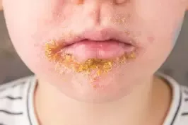
Impetigo in its most prevalent type, nonbullous.
Vesiculopustule formation that bursts results in crusting with a distinctive golden look; local lymphadenopathy may happen.
Bullous impetigo is a staphylococcal impetigo that affects the trunk more frequently and proceeds from small to large flaccid bullae (newborns/young children) as a result of the production of an epidermolytic toxin.
Folliculitis is sometimes referred to as S. aureus impetigo of the hair follicles, while Ecthyma is a more severe impetigo infection that commonly has lymphadenitis. Folliculitis affects the skin and exocrine system.
Epidemiology Incidence Males outnumber women in terms of predominance Children between the ages of 2 and 5 make up the majority. Prevalence
In the US: not documented, but widespread
Child Safety Considerations
In young children, poststreptococcal glomerulonephritis may occur after impetigo.
Impetigo neonatorum may develop as a result of pollution in nurseries.
Pathophysiology and Etiology
Pure culture of coagulase-positive staphylococci is between 50 and 90 percent; these bacteria are more contagious through touch. Pure culture of hemolytic streptococci, mostly group A, is only about 10 percent.
Mixed infections with streptococci and staphylococci are frequent; data indicate that staphylococci have become more significant over the past few decades. Methicillin-resistant S. aureus (MRSA) has been found in some cases and can spread through direct contact or insects.
Localized lymphadenopathy
Risk factors include a warm, humid environment, a tropical or subtropical climate, summer or fall, minor injuries, insect bites, and skin breaches; poor hygiene; scarcity; epidemics; war; familial transmission; poor health with anemia and malnutrition; complications from pediculosis; scabies; chickenpox; eczema/atopic dermatitis; contact dermatitis (Rhus spp. ); burns; contact sports; childcare
Prevention includes: covering wounds; paying close attention to family hygiene, especially when it comes to youngsters washing their hands; avoiding crowding and sharing of personal goods; and treating atopic dermatitis.
Malnutrition and anemia, crowded living quarters, poor cleanliness, neglected minor trauma, any chronic or underlying dermatitis, and the possibility of coinfection with scabies are all associated disorders.
Lesions are frequently reported as painful, and they may develop slowly or quickly. They are most common on the face around the mouth and nose or at the site of trauma.
clinical assessment
Early lesions include tender red papules or macules (contact dermatitis manifests as itchy lesions), thin-roofed vesicles to bullae, pustules, weeping red ulcers, honey-colored crusts, satellite lesions, frequently multiple sites, and bullae on the face, trunk, and buttocks.
Nonbullous - Contact dermatitis - Chickenpox - Herpes - Folliculitis - Erysipelas - Insect bites - Severe eczematous dermatitis - Scabies - Tinea corporis are among the differential diagnoses.
Bullous pemphigoid, Burns, Pemphigus vulgaris, and Stevens-Johnson syndrome
Initial test results from the laboratory and imaging
None are typically required in typical presentations; pus/bullae fluid cultures may be useful if empiric therapy is ineffective.
After the crust has been removed from the lesion, a culture is done, and both staphylococci and group A streptococci can develop there. - Antistreptolysin-O (ASO) titer: a weak streptococcal positive but not generally useful Antihyaluronidase (AHT) and Antideoxyribonuclease B (anti-DNase B) responses are more dependable than ASO responses. - Streptozyme: streptococci detected.
Disorders that could affect lab results include: Tests for streptococcal enzymes will be affected by streptococcal pharyngitis.
Tests in the Future & Special Considerations
In the case of impetigo with subsequent poststreptococcal glomerulonephritis, serologic testing is useful in monitoring the disease's progress and systemic symptoms.
Management
Treatment prevents illness spread, expedites recovery, and enhances cosmetic appearance.
Apply mupirocin ointment TID on the places of minor skin damage to prevent infection.
Washing the entire body may prevent recurrence at distant areas. Remove crusts, clean with gentle washing 2 to 3 times each day, and clean with antibacterial soap, chlorhexidine, or Betadine.
medicine: In 2014, the Infectious Diseases Society of America (IDSA) advised oral medicine when the condition was more severe or widespread, and topical treatment for small lesions. Topical mupirocin is similarly or even more efficacious than oral medications for treating nonextensive impetigo, according to a 2017 Canadian systematic review (2).[A]. The use of macrolides and penicillin is no longer advised. Considering the patterns of resistance, fluoroquinolones are not recommended.
● For information about microbial resistance, speak with your neighborhood hospital or health department.
Bullous (treat 10 days), nonbullous (treat 7 days), widespread (treat 10 days). - Mupirocin (Bactroban) 2% topical ointment given TID for 5–7 days only on nonbullous skin; less effective on scalp than around mouth - Retapamulin 1% ointment, which must be used BID for 5 days at a high cost. - Dicloxacillin: Adults: 250 mg PO QID; Children: 12 to 25 mg/kg/day divided every six hours; Children above 40 kg: 125 to 250 q6h.
Unless local staphylococcal strains are resistant, dicloxacillin, cephalexin, topical mupirocin, and fusidic acid are effective treatments. Tetracyclines, trimethoprim-sulfamethoxazole, or clindamycin are among the available treatments for MRSA infections. Usually, seven days of oral dosages is adequate.
Cefadroxil 30 mg/kg/day divided BID - Adults 1st-generation cephalosporins - Children Cephalexin 25 to 50 mg/kg/day divided, q6-12h Cefaclor 20 to 40 mg/kg/day divided q8h Cephradine 25 to 50 mg/kg/day divided q6-12h
Cefaclor 250 mg TID, Cephalexin 250 mg QID, and Cephradine 500 mg BID
Clindamycin 300 mg every 6 to 8 hours. Severe bullous disease may necessitate IV therapy with nafcillin or cefazolin. Cefadroxil 1 g/day in divided dosages.
Referral
If persistent infections develop, especially in people with impaired immune systems
Further Treatments
Keep an eye out for microbial resistance trends.
Athletes are prohibited from participating in contact sports. School and childcare infectious limitations are in place. Children may return to school 24 hours after the start of antimicrobial therapy.
patient observation
Culture the lesions if they don't disappear after 7 to 10 days.
Patient Education The key to preventing the transmission of infections is to practice good hand hygiene, especially when it comes to young children.
Complete remission is expected with treatment in 7 to 10 days.
Unlike rheumatic fever, antibiotic treatment will not stop or prevent glomerulonephritis.
If the matter is not resolved in 7 to 10 days, culture is required to identify the resistant bacterium.
Recurrent impetigo: Check for S. aureus carriage in the nares (as well as the perineum, axillae, and toe web). For 5 days, BID, apply mupirocin ointment to the nares for decolonization.
Ecthyma, erysipelas, post-streptococcal acute glomerulonephritis, cellulitis, bacteremia, osteomyelitis, septic arthritis, pneumonia, lymphadenitis, and lymphoma are complications.
Primary impetigo (pyoderma): invasion of previously normal skin Secondary impetigo (impetiginization): invasion at sites of minor trauma (abrasions, insect bites, underlying eczema) Infected patients typically have multiple lesions. Impetigo is a contagious, superficial, intraepidermal infection most frequently seen in children.
Staphylococcus aureus cultures are positive in >80% of cases, either by itself or in combination with group A -hemolytic streptococci; S. aureus has been the more prevalent pathogen since the 1990s.
Impetigo in its most prevalent type, nonbullous.
Vesiculopustule formation that bursts results in crusting with a distinctive golden look; local lymphadenopathy may happen.
Bullous impetigo is a staphylococcal impetigo that affects the trunk more frequently and proceeds from small to large flaccid bullae (newborns/young children) as a result of the production of an epidermolytic toxin.
Folliculitis is sometimes referred to as S. aureus impetigo of the hair follicles, while Ecthyma is a more severe impetigo infection that commonly has lymphadenitis. Folliculitis affects the skin and exocrine system.
Epidemiology Incidence Males outnumber women in terms of predominance Children between the ages of 2 and 5 make up the majority. Prevalence
In the US: not documented, but widespread
Child Safety Considerations
In young children, poststreptococcal glomerulonephritis may occur after impetigo.
Impetigo neonatorum may develop as a result of pollution in nurseries.
Pathophysiology and Etiology
Pure culture of coagulase-positive staphylococci is between 50 and 90 percent; these bacteria are more contagious through touch. Pure culture of hemolytic streptococci, mostly group A, is only about 10 percent.
Mixed infections with streptococci and staphylococci are frequent; data indicate that staphylococci have become more significant over the past few decades. Methicillin-resistant S. aureus (MRSA) has been found in some cases and can spread through direct contact or insects.
Localized lymphadenopathy
Risk factors include a warm, humid environment, a tropical or subtropical climate, summer or fall, minor injuries, insect bites, and skin breaches; poor hygiene; scarcity; epidemics; war; familial transmission; poor health with anemia and malnutrition; complications from pediculosis; scabies; chickenpox; eczema/atopic dermatitis; contact dermatitis (Rhus spp. ); burns; contact sports; childcare
Prevention includes: covering wounds; paying close attention to family hygiene, especially when it comes to youngsters washing their hands; avoiding crowding and sharing of personal goods; and treating atopic dermatitis.
Malnutrition and anemia, crowded living quarters, poor cleanliness, neglected minor trauma, any chronic or underlying dermatitis, and the possibility of coinfection with scabies are all associated disorders.
Lesions are frequently reported as painful, and they may develop slowly or quickly. They are most common on the face around the mouth and nose or at the site of trauma.
clinical assessment
Early lesions include tender red papules or macules (contact dermatitis manifests as itchy lesions), thin-roofed vesicles to bullae, pustules, weeping red ulcers, honey-colored crusts, satellite lesions, frequently multiple sites, and bullae on the face, trunk, and buttocks.
Nonbullous - Contact dermatitis - Chickenpox - Herpes - Folliculitis - Erysipelas - Insect bites - Severe eczematous dermatitis - Scabies - Tinea corporis are among the differential diagnoses.
Bullous pemphigoid, Burns, Pemphigus vulgaris, and Stevens-Johnson syndrome
Initial test results from the laboratory and imaging
None are typically required in typical presentations; pus/bullae fluid cultures may be useful if empiric therapy is ineffective.
After the crust has been removed from the lesion, a culture is done, and both staphylococci and group A streptococci can develop there. - Antistreptolysin-O (ASO) titer: a weak streptococcal positive but not generally useful Antihyaluronidase (AHT) and Antideoxyribonuclease B (anti-DNase B) responses are more dependable than ASO responses. - Streptozyme: streptococci detected.
Disorders that could affect lab results include: Tests for streptococcal enzymes will be affected by streptococcal pharyngitis.
Tests in the Future & Special Considerations
In the case of impetigo with subsequent poststreptococcal glomerulonephritis, serologic testing is useful in monitoring the disease's progress and systemic symptoms.
Management
Treatment prevents illness spread, expedites recovery, and enhances cosmetic appearance.
Apply mupirocin ointment TID on the places of minor skin damage to prevent infection.
Washing the entire body may prevent recurrence at distant areas. Remove crusts, clean with gentle washing 2 to 3 times each day, and clean with antibacterial soap, chlorhexidine, or Betadine.
medicine: In 2014, the Infectious Diseases Society of America (IDSA) advised oral medicine when the condition was more severe or widespread, and topical treatment for small lesions. Topical mupirocin is similarly or even more efficacious than oral medications for treating nonextensive impetigo, according to a 2017 Canadian systematic review (2).[A]. The use of macrolides and penicillin is no longer advised. Considering the patterns of resistance, fluoroquinolones are not recommended.
● For information about microbial resistance, speak with your neighborhood hospital or health department.
Bullous (treat 10 days), nonbullous (treat 7 days), widespread (treat 10 days). - Mupirocin (Bactroban) 2% topical ointment given TID for 5–7 days only on nonbullous skin; less effective on scalp than around mouth - Retapamulin 1% ointment, which must be used BID for 5 days at a high cost. - Dicloxacillin: Adults: 250 mg PO QID; Children: 12 to 25 mg/kg/day divided every six hours; Children above 40 kg: 125 to 250 q6h.
Unless local staphylococcal strains are resistant, dicloxacillin, cephalexin, topical mupirocin, and fusidic acid are effective treatments. Tetracyclines, trimethoprim-sulfamethoxazole, or clindamycin are among the available treatments for MRSA infections. Usually, seven days of oral dosages is adequate.
Cefadroxil 30 mg/kg/day divided BID - Adults 1st-generation cephalosporins - Children Cephalexin 25 to 50 mg/kg/day divided, q6-12h Cefaclor 20 to 40 mg/kg/day divided q8h Cephradine 25 to 50 mg/kg/day divided q6-12h
Cefaclor 250 mg TID, Cephalexin 250 mg QID, and Cephradine 500 mg BID
Clindamycin 300 mg every 6 to 8 hours. Severe bullous disease may necessitate IV therapy with nafcillin or cefazolin. Cefadroxil 1 g/day in divided dosages.
Referral
If persistent infections develop, especially in people with impaired immune systems
Further Treatments
Keep an eye out for microbial resistance trends.
Athletes are prohibited from participating in contact sports. School and childcare infectious limitations are in place. Children may return to school 24 hours after the start of antimicrobial therapy.
patient observation
Culture the lesions if they don't disappear after 7 to 10 days.
Patient Education The key to preventing the transmission of infections is to practice good hand hygiene, especially when it comes to young children.
Complete remission is expected with treatment in 7 to 10 days.
Unlike rheumatic fever, antibiotic treatment will not stop or prevent glomerulonephritis.
If the matter is not resolved in 7 to 10 days, culture is required to identify the resistant bacterium.
Recurrent impetigo: Check for S. aureus carriage in the nares (as well as the perineum, axillae, and toe web). For 5 days, BID, apply mupirocin ointment to the nares for decolonization.
Ecthyma, erysipelas, post-streptococcal acute glomerulonephritis, cellulitis, bacteremia, osteomyelitis, septic arthritis, pneumonia, lymphadenitis, and lymphoma are complications.

0 Comments
