- Published on
Pathology - Bronchiectasis
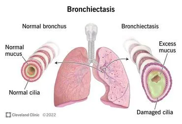
Bronchiectasis is a medical condition characterized by the abnormal widening and damage of the bronchial tubes in the lungs.
The cause of bronchial blockage can be attributed to factors such as tumors, foreign bodies, or excessive mucus. Additionally, persistent necrotizing infections of the bronchi or an underlying systemic ailment, such as cystic fibrosis (CF) or common variable immunodeficiency, can also contribute to this condition.
Lung: Enlarged air passages often found in the lower lobes; inflammation occurring inside the walls of the bronchial tubes; fibrosis of the bronchial walls in cases of chronic illness.
The patient presents with a persistent cough accompanied by a large amount of thick, pus-filled mucus. They also experience coughing up blood, bluish discoloration of the skin due to lack of oxygen, and a low red blood cell count.
Possible complications encompass the development of abscesses in the lungs and brain, as well as the occurrence of cor pulmonale.
Image capture and processing: CT scan reveals bronchioles that are enlarged and have a characteristic signet-ring look.
Laboratory results indicate a reduced ratio of forced expiratory volume to forced vital capacity (FEV/FVC) and a decreased hematocrit (Hct).
Treatment options for this condition include antibiotics, inhaled bronchodilators, inhaled steroids during exacerbations, and surgical resection for localized illness.
Kartagener syndrome arises from a dynein malfunction, resulting in cilia that are incapable of movement.
The clinical presentation encompasses bronchiectasis, infertility, recurrent sinusitis, and situs inversus (dextrocardia).
Bronchiectasis is a medical condition characterized by the abnormal widening and damage of the bronchial tubes in the lungs.
The cause of bronchial blockage can be attributed to factors such as tumors, foreign bodies, or excessive mucus. Additionally, persistent necrotizing infections of the bronchi or an underlying systemic ailment, such as cystic fibrosis (CF) or common variable immunodeficiency, can also contribute to this condition.
Lung: Enlarged air passages often found in the lower lobes; inflammation occurring inside the walls of the bronchial tubes; fibrosis of the bronchial walls in cases of chronic illness.
The patient presents with a persistent cough accompanied by a large amount of thick, pus-filled mucus. They also experience coughing up blood, bluish discoloration of the skin due to lack of oxygen, and a low red blood cell count.
Possible complications encompass the development of abscesses in the lungs and brain, as well as the occurrence of cor pulmonale.
Image capture and processing: CT scan reveals bronchioles that are enlarged and have a characteristic signet-ring look.
Laboratory results indicate a reduced ratio of forced expiratory volume to forced vital capacity (FEV/FVC) and a decreased hematocrit (Hct).
Treatment options for this condition include antibiotics, inhaled bronchodilators, inhaled steroids during exacerbations, and surgical resection for localized illness.
Kartagener syndrome arises from a dynein malfunction, resulting in cilia that are incapable of movement.
The clinical presentation encompasses bronchiectasis, infertility, recurrent sinusitis, and situs inversus (dextrocardia).
0 Comments
