- Published on
Pathology - Bullous Pemphigoid and Pemphigus Vulgaris
Bullous pemphigoid: An autoimmune condition that primarily affects males over 60 and is typified by lgG antibodies against hemidesmosomes of the epidermal basement membrane.
IgG antibodies against intercellular connections between epidermal keratinocytes are the hallmark of the autoimmune disease pemphigus vulgaris, which is most frequently observed in people between the ages of 40 and 60.
Bullous Pemphigoid: Skin has subepidermal bullae; perivascular infiltration of lymphocytes and eosinophils; immunofluorescence shows IgG deposition and a complement band running along the basement membrane.
In Pemphigus vulgaris, cells just above the skin's basal cell layer undergo acantholysis, resulting in a suprabasal acantholytic blister; immunofluorescence shows that complement and IgG surround epidermal cells.
Clinically less severe than pemphigus vulgaris, bullous pemphigoid is a chronic, relapsing, and remitting condition characterized by pruritic, fluid-filled blisters. Antibasement membrane antibodies were found in the lab.
Pneumonia pemphigus vulgaris: severe intraepidermal bullae that start in the lips and spread throughout the body; bullae ruptures can result in secondary infections; positive Nikolsky sign (blister formation following finger rubbing). Immunoglobulins, an antiadhesion molecule, were found in the lab.
Systemic corticosteroids for bullous pemphigoid; other immunosuppressive medications may be needed in more severe instances.
Systemic corticosteroids and immunosuppressive medications (such mycophenolate mofetil) are used to treat Pemphigus vulgaris.
In contrast to PV, paraneoplastic pemphigus is linked to mucosal lesions, just like EM. Because of the underlying malignancy, survival rates are dismal.
Bullous pemphigoid: An autoimmune condition that primarily affects males over 60 and is typified by lgG antibodies against hemidesmosomes of the epidermal basement membrane.
IgG antibodies against intercellular connections between epidermal keratinocytes are the hallmark of the autoimmune disease pemphigus vulgaris, which is most frequently observed in people between the ages of 40 and 60.
Bullous Pemphigoid: Skin has subepidermal bullae; perivascular infiltration of lymphocytes and eosinophils; immunofluorescence shows IgG deposition and a complement band running along the basement membrane.
In Pemphigus vulgaris, cells just above the skin's basal cell layer undergo acantholysis, resulting in a suprabasal acantholytic blister; immunofluorescence shows that complement and IgG surround epidermal cells.
Clinically less severe than pemphigus vulgaris, bullous pemphigoid is a chronic, relapsing, and remitting condition characterized by pruritic, fluid-filled blisters. Antibasement membrane antibodies were found in the lab.
Pneumonia pemphigus vulgaris: severe intraepidermal bullae that start in the lips and spread throughout the body; bullae ruptures can result in secondary infections; positive Nikolsky sign (blister formation following finger rubbing). Immunoglobulins, an antiadhesion molecule, were found in the lab.
Systemic corticosteroids for bullous pemphigoid; other immunosuppressive medications may be needed in more severe instances.
Systemic corticosteroids and immunosuppressive medications (such mycophenolate mofetil) are used to treat Pemphigus vulgaris.
In contrast to PV, paraneoplastic pemphigus is linked to mucosal lesions, just like EM. Because of the underlying malignancy, survival rates are dismal.
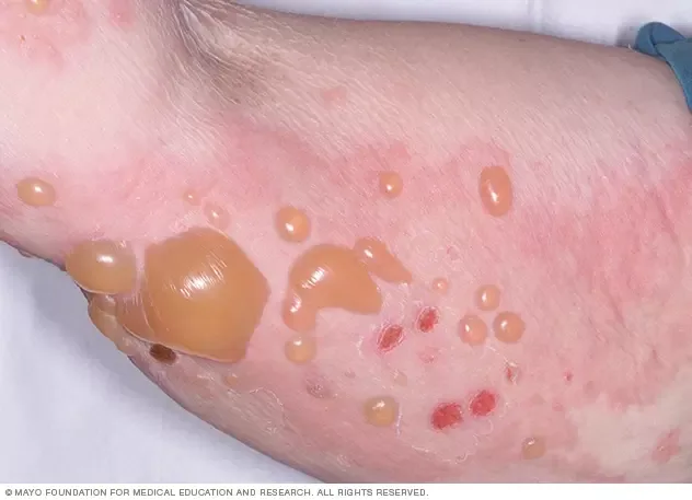
Bullous Pemphigoid
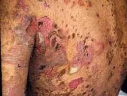
Pemphigoid Vulgaris
0 Comments
