- Published on
Pathology - Cardiopulmonary Arrest
Pathophysiology
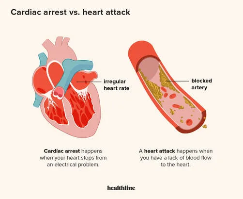
This illness is brought on by an abrupt blockage of a coronary artery, which causes an instantaneous ischemia of the heart tissue that the artery supplies. Electrolyte anomalies may change the usual membrane potential. Myocardial cells that are hypoxic undergo hyperautomaticity, in which many cells function as pacemakers. While hypoxic tissue is excitable and will contract erratically, scarred and dying tissue will not react to this electrical stimulation.
Ventricular fibrillation, which is the outcome of myocardial ischemia, is characterized by an irregular contraction of the cardiac muscle, which causes the tissue to quiver instead of the ventricles contracting in an orderly manner. A short while later, this unproductive rhythm gave way to asystole, or the lack of heart contraction.
The patient's ECG abnormalities (ischemia in leads V2-V6) points to a possible acute blockage of the left anterior descending artery, which delivers blood to the left ventricle's anterior surface. Reduced blood pressure in the context of an elevated pulse rate and low left ventricular ejection fraction is indicative of a markedly diminished cardiac output brought on by an uncoordinated and inefficient left ventricle contraction.
Pathophysiology
This illness is brought on by an abrupt blockage of a coronary artery, which causes an instantaneous ischemia of the heart tissue that the artery supplies. Electrolyte anomalies may change the usual membrane potential. Myocardial cells that are hypoxic undergo hyperautomaticity, in which many cells function as pacemakers. While hypoxic tissue is excitable and will contract erratically, scarred and dying tissue will not react to this electrical stimulation.
Ventricular fibrillation, which is the outcome of myocardial ischemia, is characterized by an irregular contraction of the cardiac muscle, which causes the tissue to quiver instead of the ventricles contracting in an orderly manner. A short while later, this unproductive rhythm gave way to asystole, or the lack of heart contraction.
The patient's ECG abnormalities (ischemia in leads V2-V6) points to a possible acute blockage of the left anterior descending artery, which delivers blood to the left ventricle's anterior surface. Reduced blood pressure in the context of an elevated pulse rate and low left ventricular ejection fraction is indicative of a markedly diminished cardiac output brought on by an uncoordinated and inefficient left ventricle contraction.
0 Comments
