- Published on
Pathology - Gout
Caused by urate crystals that are deposited together as a result of hyperuricemia.
Primary: Occurs most frequently in middle-aged males and Pacific Islanders; caused by idiopathic hyperuricemia; risk factors include obesity, alcohol consumption, and hereditary vulnerability.
Secondary: Results from hyperuricemia brought on by underlying conditions such Lesch-Nyhan syndrome (HGPRT deficiency), myeloproliferative diseases, reduced urate excretion (renal illness), and medications.
Joint: Emejected synovium with inflammatory infiltrate; urate crystals (needle-shaped, negatively birefringent crystals) and neutrophils in synovial fluid; urate deposits can eventually cause cartilage degradation.
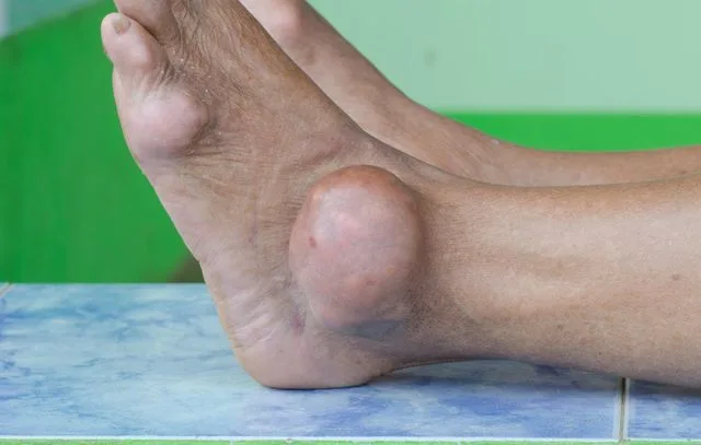
Tophi: Urate crystal cluster encircled by large cells, lymphocytes, and fibroblasts found in cartilage or soft tissues.
Acute arthritis may give way to chronic arthritis; tophi on the hands, feet, or ears may appear months after acute arthritis; urate nephropathy with interstitial deposit of urate crystals and obstruction by uric acid stones; swollen, tender joint with abrupt onset, frequently in MTP joint of big toe (podagra), foot, ankles, or knees.
Results from the lab: leukocytosis, elevated ESR, and hyperuricemia.
Treatments: probenecid or allopurinol for long-term care; colchicine, NSAIDs, and/or steroids for acute flare-ups.
Calcium pyrophosphate crystals (positive, birefringent, rhomboid crystals) deposited in joints are the source of pseudogout. Large joints, such as the knee, shoulders, and wrists, are typically affected, and it is more common among the elderly. NSAIDs or intra-articular steroids are used to treat it.
0 Comments
