- Published on
Pathology - Hydatidiform Mole and Gestational Choriocarcinoma
Hydatidiform mole (HM) occurs as a result of embryonic chromosomal anomalies. A complete mole has a chromosomal makeup of 46, XX, with all chromosomes originating from the sperm. A partial mole is triploid (69, XXY) or tetraploid, typically resulting from the fertilization of an ovum by several sperm.
Gestational choriocarcinoma (GC) is more common in Asia and Africa and is typically preceded by a complete hydatidiform mole, abortions, or ectopic or normal pregnancies.
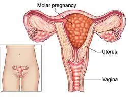
HM: Features include edematous chorionic villi with cystic swelling forming grapelike clusters, hyperplasia of trophoblastic cells, honeycombed appearance of the uterus, and lack of fetal components. Partial findings include swollen villi and the presence of embryonic body components.
GC :A tumor including necrotic and hemorrhagic patches made up of cells resembling syncytiotrophoblasts and cytotrophoblasts, with potential endometrial penetration.
HM: may manifest as nausea and vomiting, painless vaginal bleeding typically occurring in the first trimester, enlarged uterus for gestational age, and maybe hypertension in the first trimester.
Ultrasound shows a snowstorm look without detection of fetal or placental components.
GC:Excessive vaginal bleeding following the removal of a mole or after giving birth/miscarriage; hemoptysis caused by early spread of the condition to the lungs through the blood.
Laboratory results: Elevated serum hCG levels for the stage of pregnancy (HM and GC).
Performing immediate uterine evacuation via suction.
GC: Surgical excision; chemotherapy.
Hydatidiform mole (HM) occurs as a result of embryonic chromosomal anomalies. A complete mole has a chromosomal makeup of 46, XX, with all chromosomes originating from the sperm. A partial mole is triploid (69, XXY) or tetraploid, typically resulting from the fertilization of an ovum by several sperm.
Gestational choriocarcinoma (GC) is more common in Asia and Africa and is typically preceded by a complete hydatidiform mole, abortions, or ectopic or normal pregnancies.
HM: Features include edematous chorionic villi with cystic swelling forming grapelike clusters, hyperplasia of trophoblastic cells, honeycombed appearance of the uterus, and lack of fetal components. Partial findings include swollen villi and the presence of embryonic body components.
GC :A tumor including necrotic and hemorrhagic patches made up of cells resembling syncytiotrophoblasts and cytotrophoblasts, with potential endometrial penetration.
HM: may manifest as nausea and vomiting, painless vaginal bleeding typically occurring in the first trimester, enlarged uterus for gestational age, and maybe hypertension in the first trimester.
Ultrasound shows a snowstorm look without detection of fetal or placental components.
GC:Excessive vaginal bleeding following the removal of a mole or after giving birth/miscarriage; hemoptysis caused by early spread of the condition to the lungs through the blood.
Laboratory results: Elevated serum hCG levels for the stage of pregnancy (HM and GC).
Performing immediate uterine evacuation via suction.
GC: Surgical excision; chemotherapy.
0 Comments
