- Published on
Pathology - Ischemic Stroke
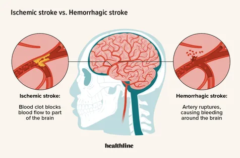
Possible causes are thrombosis, embolism, dissection, vasculitis, or hypotension.
Risk factors including tobacco use, hypertension, atrial fibrillation, hyperlipidemia, and diabetes.
Cerebral infarction is linked to thrombosis or embolism and results in ischemic neuronal changes such as nuclear pyknosis and eosinophilic cytoplasm within 12 hours. Microglia and monocyte infiltration occur within 2 days, followed by liquefactive necrosis leading to a fluid-filled cavity and reactive astrocytes within 1-3 weeks. Gliosis, which is scar formation, can occur after several months and the infarction may progress to hemorrhagic infarction.
Watershed infarction is linked to low blood pressure and manifests as a wedge-shaped infarction at the border of the artery's supply area, typically found between the distribution of the anterior cerebral artery (ACA) and middle cerebral artery (MCA).
Lacunar infarcts are linked to high blood pressure and blockage of small blood vessels due to blood clots, resulting in small empty spaces surrounded by scar tissue in the brain.
It varies based on the location of reduced blood flow and the degree of alternative blood supply.
Anterior Cerebral Artery (ACA): Contralateral leg weakness and sensory loss.
Middle Cerebral Artery (MCA) presents with contralateral paralysis, sensory loss, contralateral hemianopia, and aphasias.
Proximal Cerebral Artery (PCA) symptoms include contralateral sensory disruption and homonymous hemianopia, with or without macular-sparing.
Damage to the lateral striate arteries results in contralateral paralysis.
Administer thrombolytic therapy after 4.5 hours of symptom onset if no signs of hemorrhage. Use antiplatelet medication such as aspirin and dipyridamole. Modify risk factors by treating hypertension and hyperlipidemia.
A transient ischemic attack is a temporary episode of neurological symptoms resulting from reduced blood flow to the brain, which typically lasts less than 24 hours and indicates a high risk of a future stroke.
Possible causes are thrombosis, embolism, dissection, vasculitis, or hypotension.
Risk factors including tobacco use, hypertension, atrial fibrillation, hyperlipidemia, and diabetes.
Cerebral infarction is linked to thrombosis or embolism and results in ischemic neuronal changes such as nuclear pyknosis and eosinophilic cytoplasm within 12 hours. Microglia and monocyte infiltration occur within 2 days, followed by liquefactive necrosis leading to a fluid-filled cavity and reactive astrocytes within 1-3 weeks. Gliosis, which is scar formation, can occur after several months and the infarction may progress to hemorrhagic infarction.
Watershed infarction is linked to low blood pressure and manifests as a wedge-shaped infarction at the border of the artery's supply area, typically found between the distribution of the anterior cerebral artery (ACA) and middle cerebral artery (MCA).
Lacunar infarcts are linked to high blood pressure and blockage of small blood vessels due to blood clots, resulting in small empty spaces surrounded by scar tissue in the brain.
It varies based on the location of reduced blood flow and the degree of alternative blood supply.
Anterior Cerebral Artery (ACA): Contralateral leg weakness and sensory loss.
Middle Cerebral Artery (MCA) presents with contralateral paralysis, sensory loss, contralateral hemianopia, and aphasias.
Proximal Cerebral Artery (PCA) symptoms include contralateral sensory disruption and homonymous hemianopia, with or without macular-sparing.
Damage to the lateral striate arteries results in contralateral paralysis.
Administer thrombolytic therapy after 4.5 hours of symptom onset if no signs of hemorrhage. Use antiplatelet medication such as aspirin and dipyridamole. Modify risk factors by treating hypertension and hyperlipidemia.
A transient ischemic attack is a temporary episode of neurological symptoms resulting from reduced blood flow to the brain, which typically lasts less than 24 hours and indicates a high risk of a future stroke.
0 Comments
