- Published on
Pathology - Left Ventricular Failure
Pathophysiology
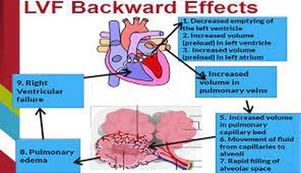
Paroxysmal nocturnal dyspnea (PND), brought on by acute pulmonary edema, is the presenting complaint. PND usually develops two to three hours during sleep and is caused by the fluid from peripheral edema entering the bloodstream when the patient is flat. Tachypnea is brought on by pulmonary edema, which also reduces lung compliance and activates mechanoreceptors. Reduced cardiac output results in inadequate tissue perfusion, as shown by chilly skin and inadequate capillary refill. Low cardiac output lowers systemic arterial blood pressure, which triggers the sympathetic nervous system (SNS) and results in tachycardia and vasoconstriction.
Lack of heat dissipation and increased metabolic heat production from sympathetic activation raise body temperature. The S3 gallop is brought on by LV hypertrophy, a persistent physiological adjustment, which fills a noncompliant ventricle with blood during early diastole. A loud P2 sound indicates the onset of pulmonary hypertension due to a rise in pressure across the pulmonic valve. On the other hand, the lack of jugular venous distension indicates that the right ventricle is still functioning.
Chronic low effective circulation volume causes the renin-angiotensin-aldosterone and vasopressin axes to be activated, which is known as neurohormonal compensation. In an unsuccessful attempt to revive cardiac output, ECF is held in reserve.
Excessive water retention causes hyponatremia, whereas renal potassium losses cause hypokalemia in this chronic secondary hyperaldosteronism condition.
Pathophysiology
Paroxysmal nocturnal dyspnea (PND), brought on by acute pulmonary edema, is the presenting complaint. PND usually develops two to three hours during sleep and is caused by the fluid from peripheral edema entering the bloodstream when the patient is flat. Tachypnea is brought on by pulmonary edema, which also reduces lung compliance and activates mechanoreceptors. Reduced cardiac output results in inadequate tissue perfusion, as shown by chilly skin and inadequate capillary refill. Low cardiac output lowers systemic arterial blood pressure, which triggers the sympathetic nervous system (SNS) and results in tachycardia and vasoconstriction.
Lack of heat dissipation and increased metabolic heat production from sympathetic activation raise body temperature. The S3 gallop is brought on by LV hypertrophy, a persistent physiological adjustment, which fills a noncompliant ventricle with blood during early diastole. A loud P2 sound indicates the onset of pulmonary hypertension due to a rise in pressure across the pulmonic valve. On the other hand, the lack of jugular venous distension indicates that the right ventricle is still functioning.
Chronic low effective circulation volume causes the renin-angiotensin-aldosterone and vasopressin axes to be activated, which is known as neurohormonal compensation. In an unsuccessful attempt to revive cardiac output, ECF is held in reserve.
Excessive water retention causes hyponatremia, whereas renal potassium losses cause hypokalemia in this chronic secondary hyperaldosteronism condition.
0 Comments
