- Published on
Pathology - Primary Focal Segmental Glomerulosclerosis
Pathophysiology
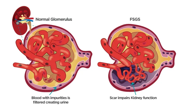
Has been linked to nephrotic syndrome. Because of the high rate of urine protein loss, alterations in capillary Starling forces result in hypoalbuminemia and the production of edema. This patient has primary (idiopathic) focal segmental glomerulosclerosis (FSGS), a condition in which podocyte abnormalities and glomerular filtration barrier failure are caused by circulating immunological stimuli or genetic disorders. The high rate of progression to end-stage renal disease makes the FSGS damage pattern observed on biopsy significant. Numerous secondary causes of FSGS exist, such as infections, nephrotoxins, cancers, and systemic disorders. The patient's medical history excludes a few typical secondary causes. "Membranous nephropathy" (MN), in which the glomerular basement membrane is observed to be thicker on light microscopy and immunostaining would reveal IgG deposition throughout the glomerular capillary loop, is another common appearance on renal biopsy of patients with nephrotic syndrome. Electron microscopy in the instance of MN would demonstrate that the thickening of the basement membrane was caused by sub-epithelial deposits and that the podocyte foot processes had been visibly effaced. MN may arise from multiple secondary causes or from a primary autoimmune disease.
Nonimmunotherapies such a low-salt diet, diuretics to alleviate edema, ACE inhibitors to lower intraglomerular blood pressure (by dilatation of efferent arteriole), and lipid-lowering medications are generally used in the treatment of nephrotic syndrome.
When MN is present, immunosuppression may also be employed.
Pathophysiology
Has been linked to nephrotic syndrome. Because of the high rate of urine protein loss, alterations in capillary Starling forces result in hypoalbuminemia and the production of edema. This patient has primary (idiopathic) focal segmental glomerulosclerosis (FSGS), a condition in which podocyte abnormalities and glomerular filtration barrier failure are caused by circulating immunological stimuli or genetic disorders. The high rate of progression to end-stage renal disease makes the FSGS damage pattern observed on biopsy significant. Numerous secondary causes of FSGS exist, such as infections, nephrotoxins, cancers, and systemic disorders. The patient's medical history excludes a few typical secondary causes. "Membranous nephropathy" (MN), in which the glomerular basement membrane is observed to be thicker on light microscopy and immunostaining would reveal IgG deposition throughout the glomerular capillary loop, is another common appearance on renal biopsy of patients with nephrotic syndrome. Electron microscopy in the instance of MN would demonstrate that the thickening of the basement membrane was caused by sub-epithelial deposits and that the podocyte foot processes had been visibly effaced. MN may arise from multiple secondary causes or from a primary autoimmune disease.
Nonimmunotherapies such a low-salt diet, diuretics to alleviate edema, ACE inhibitors to lower intraglomerular blood pressure (by dilatation of efferent arteriole), and lipid-lowering medications are generally used in the treatment of nephrotic syndrome.
When MN is present, immunosuppression may also be employed.
0 Comments
