- Published on
Pathology - Pulmonary Edema
Pathophysiology
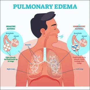
An accumulation of fluid within the lung parenchyma causes this disease. This usually happens as a consequence of uncontrolled left-sided cardiac failure. Increased LV and atrial diastolic pressure, which is then conveyed to the pulmonary veins and capillaries, is the outcome of venous return into a malfunctioning left heart.
Edema forms as a result of elevated hydrostatic pressure in pulmonary capillaries.
Because pulmonary edema activates mechanosensitive receptors in the airways, it results in reflex tachypnea and coughing. An excessive amount of interstitial fluid reduces lung compliance, which interferes with regular breathing and oxygenation. Pulmonary edema is correlated with physical examination findings of rales (crackles, crepitations) on auscultation. The SNS is activated in tachycardia as a result of anxiety associated to the patient's dyspnea experience. Pulmonary infiltrates are typically seen on a chest radiograph, mostly in the medial and basilar lung areas.
Uncompensated left-sided heart failure can result in pulmonary edema and pleural effusions. Diuretic drugs are sometimes used to treat heart failure patients' pulmonary edema; nevertheless, stopping these drugs suddenly can make conditions worse, as in this instance.
Pathophysiology
An accumulation of fluid within the lung parenchyma causes this disease. This usually happens as a consequence of uncontrolled left-sided cardiac failure. Increased LV and atrial diastolic pressure, which is then conveyed to the pulmonary veins and capillaries, is the outcome of venous return into a malfunctioning left heart.
Edema forms as a result of elevated hydrostatic pressure in pulmonary capillaries.
Because pulmonary edema activates mechanosensitive receptors in the airways, it results in reflex tachypnea and coughing. An excessive amount of interstitial fluid reduces lung compliance, which interferes with regular breathing and oxygenation. Pulmonary edema is correlated with physical examination findings of rales (crackles, crepitations) on auscultation. The SNS is activated in tachycardia as a result of anxiety associated to the patient's dyspnea experience. Pulmonary infiltrates are typically seen on a chest radiograph, mostly in the medial and basilar lung areas.
Uncompensated left-sided heart failure can result in pulmonary edema and pleural effusions. Diuretic drugs are sometimes used to treat heart failure patients' pulmonary edema; nevertheless, stopping these drugs suddenly can make conditions worse, as in this instance.
0 Comments
