- Published on
Pathology - Rickets and Osteomalacia
caused by a lack of vitamin D, which can be brought on by starvation, insufficient exposure to sunshine, fat malabsorption syndromes (induced by pancreatic insufficiency, liver illness, inflammatory bowel disease), or kidney disease (caused by a malfunction in the kidneys' ability to synthesise l,25(0H2)D).
Aluminum toxicity, phosphate shortage, and calcium deficit in the diet are additional factors.
The condition in children is called rickets, while the sickness in adults is called osteomalacia.
Pathophysiology: Lack of vitamin D reduced the kidney's and intestines' ability to absorb Ca2+, which in turn reduced the calcification of the osteoid matrix.
Increased unmineralized bone matrix in the bone; trabeculae contain a calcified bone core encircled by an unmineralized osteoid sheath.
Clinical Signs and Symptoms
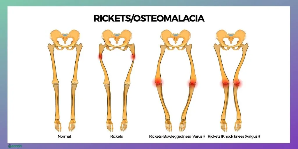
Rickets symptoms include reduced stature, rachitic rosary (thickening of the costochondral junction), pigeon breast (protrusion of the sternum), late fontanelle closure, and craniotabes (thinned occipital and parietal bones).
Osteomalacia: Weakness in the muscles and diffuse bone discomfort. Imaging: A radiograph displaying diffuse radiolucency and thinning cortical bone.
Results from the laboratory for osteomalacia and rickets: reduced phosphate and calcium concentrations.
Treatments
supplementation of vitamin D; underlying cause therapy.
Renal osteodystrophy is the term used to describe osteomalacia caused by chronic renal illness that leads in a relative vitamin D shortage and secondary hyperparathyroidism.
caused by a lack of vitamin D, which can be brought on by starvation, insufficient exposure to sunshine, fat malabsorption syndromes (induced by pancreatic insufficiency, liver illness, inflammatory bowel disease), or kidney disease (caused by a malfunction in the kidneys' ability to synthesise l,25(0H2)D).
Aluminum toxicity, phosphate shortage, and calcium deficit in the diet are additional factors.
The condition in children is called rickets, while the sickness in adults is called osteomalacia.
Pathophysiology: Lack of vitamin D reduced the kidney's and intestines' ability to absorb Ca2+, which in turn reduced the calcification of the osteoid matrix.
Increased unmineralized bone matrix in the bone; trabeculae contain a calcified bone core encircled by an unmineralized osteoid sheath.
Clinical Signs and Symptoms
Rickets symptoms include reduced stature, rachitic rosary (thickening of the costochondral junction), pigeon breast (protrusion of the sternum), late fontanelle closure, and craniotabes (thinned occipital and parietal bones).
Osteomalacia: Weakness in the muscles and diffuse bone discomfort. Imaging: A radiograph displaying diffuse radiolucency and thinning cortical bone.
Results from the laboratory for osteomalacia and rickets: reduced phosphate and calcium concentrations.
Treatments
supplementation of vitamin D; underlying cause therapy.
Renal osteodystrophy is the term used to describe osteomalacia caused by chronic renal illness that leads in a relative vitamin D shortage and secondary hyperparathyroidism.
0 Comments
