- Published on
Pathology -Systemic Lupus Erythematosus
Autoimmune disorder; linked to HLA-DR2 and -DR3; some medications (phenytoin, procainamide, INH, hydralazine) can cause a syndrome that is reversible and resembles systemic lupus erythematosus (SLE).
African American women between the ages of 20 and 40 are most frequently affected.
Pathophysiology: The disease is caused by either autoantibody-mediated host cell death or a type III hypersensitivity reaction resulting in the deposition of antigen-antibody complexes in the capillaries of visceral tissues.
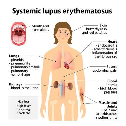
Heart: Myocarditis, pericarditis, and mitral valve disease; early coronary artery disease may also be present.
Kidney: Immune complex deposition and wire-loop lesions.
Skin: Necrotizing vasculitis; destruction of the epidermal basal layer accompanied by cutaneous edema.
Joint: synovitis with mononuclear infiltration in subsynovial tissue and neutrophils in synovial fluid.
Lung: interstitial fibrosis, pleuritis, and pleural effusions.
Clinical Signs and Symptoms
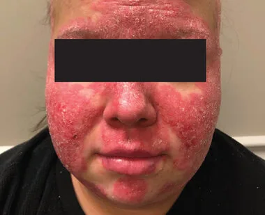
fever, exhaustion, rash resembling a butterfly, hair loss, mucosal ulcers, arthritis, photosensitivity, convulsions or amnesia, pleuritis; kidney disease with proteinuria, Raynaud phenomenon, and Libman-Sacks endocarditis.
Results from the lab: Possession of positive ANA, anti-ds DNA, and/or anti-Smith antibodies; possible detection of antihistone antibodies in drug-induced SLE; erroneous positive results on the RPR/VDRL syphilis test; hemolytic anemia; pancytopenia.
NSAIDs, corticosteroids, and immunomodulators (such as cyclophosphamide and mycophenolate mofetil) are used as treatments.
Autoimmune disorder; linked to HLA-DR2 and -DR3; some medications (phenytoin, procainamide, INH, hydralazine) can cause a syndrome that is reversible and resembles systemic lupus erythematosus (SLE).
African American women between the ages of 20 and 40 are most frequently affected.
Pathophysiology: The disease is caused by either autoantibody-mediated host cell death or a type III hypersensitivity reaction resulting in the deposition of antigen-antibody complexes in the capillaries of visceral tissues.
Heart: Myocarditis, pericarditis, and mitral valve disease; early coronary artery disease may also be present.
Kidney: Immune complex deposition and wire-loop lesions.
Skin: Necrotizing vasculitis; destruction of the epidermal basal layer accompanied by cutaneous edema.
Joint: synovitis with mononuclear infiltration in subsynovial tissue and neutrophils in synovial fluid.
Lung: interstitial fibrosis, pleuritis, and pleural effusions.
Clinical Signs and Symptoms
fever, exhaustion, rash resembling a butterfly, hair loss, mucosal ulcers, arthritis, photosensitivity, convulsions or amnesia, pleuritis; kidney disease with proteinuria, Raynaud phenomenon, and Libman-Sacks endocarditis.
Results from the lab: Possession of positive ANA, anti-ds DNA, and/or anti-Smith antibodies; possible detection of antihistone antibodies in drug-induced SLE; erroneous positive results on the RPR/VDRL syphilis test; hemolytic anemia; pancytopenia.
NSAIDs, corticosteroids, and immunomodulators (such as cyclophosphamide and mycophenolate mofetil) are used as treatments.
0 Comments
