- Published on
Surgery - Above Knee Amputation
Indications
gangrene, ischemia, or infarction: lower limb ischaemia, whether acute or chronic, brought on by severe burns or trauma.
Cancer: certain tumors, such as osteosarcoma.
Severe infection: necrotizing fasciitis or gas gangrene (Clostridium perfringens).
Unmanageable ulcers or excruciatingly paralyzed limbs are uncommon.
Anatomy
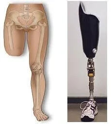
Above Knee Amputation: It is ideal to be 15 cm above the tibial plateau.
Through-knee amputation: In some cases, this is necessary (for example, if the femur has previously undergone orthopaedic stabilization). The drawbacks include erratic skin flap healing and a bulbous stump that requires prosthesis fitting.
A lengthier stump than an AKA is left behind after a femur division at the supracondylar level, providing support for the patient during sitting.
Other procedures (such as amputation of the hindquarter and disarticulation of the hip) are infrequent and are primarily done for severe infections or cancer.
Investigational studies
Pre-operative care should ideally involve a multidisciplinary evaluation by prosthetic, anesthetic, and surgical specialists. evaluation of the degree of amputation in light of the patient's characteristics and the severity of the condition (e.g. rehabilitation chances). If a patient has diabetes, a sliding insulin scale, proper blood tests and cross-matching, and, if necessary, urine catheterization.
Following surgery: Early physiotherapy, prosthesis fitting, or walking aids (such as a pneumatic postamputation mobility device) are used in the rehabilitation process.
Actions
Access: The skin is marked by two identical fish mouth-shaped skin flaps, the upper ends of which are located at the level of the femur transaction. 15 cm above the tibial plateau is this location.
Ligation of muscles and vessels: During skin incision, diathermy is used to split the muscles of the anterior and posterior thigh compartments and ligate the long saphenous vein.
Sutures connect the quadriceps to the hamstrings and the vastus lateralis to the adductors. Nerves are neatly split under mild traction, and arteries and veins are tied off.
A bone amputation involves dividing the femur, removing the periosteum, and polishing the ends of the bone to smooth it out.
Closure: After achieving hemostasis, the skin is sutured together by bringing the two myoplastic flaps together. You might leave a drain in place.
Complications
Early: Bony spurs, psychological issues, stump length that is too long or short, DVT, flap ischaemia, stump hemorrhage, neuroma, or infection; 15% early mortality.
Late: "Phantom" limb pain (lessened by potent analgesics after surgery), neuroma development, skin-borne bone erosion, ischaemia, osteomyelitis, ulceration.
Prognosis
The majority of people who have amputations have concurrent severe atherosclerotic disease, and only 30% of them survive five years after the procedure due to a significant risk of further vascular complications.
Indications
gangrene, ischemia, or infarction: lower limb ischaemia, whether acute or chronic, brought on by severe burns or trauma.
Cancer: certain tumors, such as osteosarcoma.
Severe infection: necrotizing fasciitis or gas gangrene (Clostridium perfringens).
Unmanageable ulcers or excruciatingly paralyzed limbs are uncommon.
Anatomy
Above Knee Amputation: It is ideal to be 15 cm above the tibial plateau.
Through-knee amputation: In some cases, this is necessary (for example, if the femur has previously undergone orthopaedic stabilization). The drawbacks include erratic skin flap healing and a bulbous stump that requires prosthesis fitting.
A lengthier stump than an AKA is left behind after a femur division at the supracondylar level, providing support for the patient during sitting.
Other procedures (such as amputation of the hindquarter and disarticulation of the hip) are infrequent and are primarily done for severe infections or cancer.
Investigational studies
Pre-operative care should ideally involve a multidisciplinary evaluation by prosthetic, anesthetic, and surgical specialists. evaluation of the degree of amputation in light of the patient's characteristics and the severity of the condition (e.g. rehabilitation chances). If a patient has diabetes, a sliding insulin scale, proper blood tests and cross-matching, and, if necessary, urine catheterization.
Following surgery: Early physiotherapy, prosthesis fitting, or walking aids (such as a pneumatic postamputation mobility device) are used in the rehabilitation process.
Actions
Access: The skin is marked by two identical fish mouth-shaped skin flaps, the upper ends of which are located at the level of the femur transaction. 15 cm above the tibial plateau is this location.
Ligation of muscles and vessels: During skin incision, diathermy is used to split the muscles of the anterior and posterior thigh compartments and ligate the long saphenous vein.
Sutures connect the quadriceps to the hamstrings and the vastus lateralis to the adductors. Nerves are neatly split under mild traction, and arteries and veins are tied off.
A bone amputation involves dividing the femur, removing the periosteum, and polishing the ends of the bone to smooth it out.
Closure: After achieving hemostasis, the skin is sutured together by bringing the two myoplastic flaps together. You might leave a drain in place.
Complications
Early: Bony spurs, psychological issues, stump length that is too long or short, DVT, flap ischaemia, stump hemorrhage, neuroma, or infection; 15% early mortality.
Late: "Phantom" limb pain (lessened by potent analgesics after surgery), neuroma development, skin-borne bone erosion, ischaemia, osteomyelitis, ulceration.
Prognosis
The majority of people who have amputations have concurrent severe atherosclerotic disease, and only 30% of them survive five years after the procedure due to a significant risk of further vascular complications.
0 Comments
