- Published on
Surgery - Anal Carcinoma
Introduction
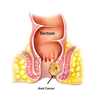
Malignancy developing in the anal verge or canal.
R isk Factors
connected to human papillomavirus carcinogenic strains (e.g. 16, 18). older than fifty. HIV or immunosuppressive medications, chronic fistulae, prior pelvic irradiation, genital warts, several sexual partners, homosexual men, and those having anorexic relations may further increase risk, while the increased risk decreases with quitting smoking.
Epidemiology
Rare, 3–4% of large bowel carcinomas, mean age 50–70 years, females >males (although anal border tumors are more prevalent in men). increasing frequency.
H HISTORY
Anal discomfort, bleeding, pruritus, discharge, tenesmus, and a mass felt on the anal edge. faecal incontinence if sphincter involvement. Absence of symptoms.
Examination
An examination of the anal edge may reveal an ulcer or proliferative growth, while a PR examination may reveal a region of inflammation or mass. At presentation, 15–30% of patients will have palpable inguinal lymph nodes (albeit only 50% of them will have tumors).
Pathogenesis
The most common kind, which is typically unpigmented, is squamous cell carcinoma (80%), adenocarcinoma, or infrequently malignant melanoma (the most common place after skin and eye). Dysplasia of the squamous epithelium is referred to as anal intraepithelial neoplasia, and it is believed to be pre-malignant.
Anal cancer is categorized according to its location:. Anal canal tumors are typically non-keratinizing, poorly differentiated tumors. 15% to 30% of anal margin tumors are often well differentiated and produce keratin.
While tumors below the dentate line spread to the inguinal nodes, tumors above the dentate line spread to the pelvic lymph nodes.
Investigational studies
For histology, proctoscopy and biopsy are used. It can be necessary to do an examination while sedated.
Blood: LFT, FBC (for anemia).
Imaging: PET scan for staging, MRI and CT, endoanal ultrasonography to evaluate invasion.
TNM staging scheme: T0: no indication of a primary tumor That is: in situ carcinoma; TI: 2 cm, T2: 2 cm, < 5 cm; T3 > 5 cm; T4: any size but is expanding into the surrounding tissues. N0 denotes the absence of lymph nodes, N1 the involvement of perirectal lymph nodes, N2 the involvement of unilateral pelvic or inguinal nodes, and N3 the involvement of bilateral pelvic or groin nodes or perirectal þ pelvis or groin nodes. Nx: Unassessed regional nodes. M0: no distant spread; M1: metastases to distant organs or abdominal lymph nodes; Mx: metastasis not assessed.
Management
Prevention: Reduce the amount of risk. A vaccination against HPV 16 and 18 is available, however as of right now, it can only be used to prevent cervical cancer. There are plans for more research on the vaccine's potential to prevent anal cancer.
Radiation and chemotherapy: In addition to local irradiation, agents such as 5-fluorouracil and mitomicinCor cisplatin are administered to the inguinal nodes and anal region.
Results similar to radical surgery where the anal sphincter is preserved
Surgery: Local excision of anal border small epidermoid carcinomas may be therapeutic.
Abdominoperineal resection was previously used to treat carcinomas of the anal canal; however, due to advancements in chemoradiotherapy, this procedure is now typically reserved for patients with residual disease, recurrence following radiotherapy, obstructive cancers, or other anal canal malignancies (such as adenocarcinoma).
Complications
Local: If ignored, there may be rectovaginal fistula, pain, bleeding, and incontinence.
Regarding radiotherapy: diarrhea, proctitis, dermatitis, and irritation of the perineum brought on by radiation.
Prognosis
Histological type, location, differentiation, and stage are significant variables. When severe chemotherapy and radiation are used to treat early-stage squamous carcinomas, the five-year survival rate drops to 30% if inguinal nodes are implicated. In this area, the prognosis for melanoma is poor, with just 10% of cases being cured with surgery.
Introduction
Malignancy developing in the anal verge or canal.
R isk Factors
connected to human papillomavirus carcinogenic strains (e.g. 16, 18). older than fifty. HIV or immunosuppressive medications, chronic fistulae, prior pelvic irradiation, genital warts, several sexual partners, homosexual men, and those having anorexic relations may further increase risk, while the increased risk decreases with quitting smoking.
Epidemiology
Rare, 3–4% of large bowel carcinomas, mean age 50–70 years, females >males (although anal border tumors are more prevalent in men). increasing frequency.
H HISTORY
Anal discomfort, bleeding, pruritus, discharge, tenesmus, and a mass felt on the anal edge. faecal incontinence if sphincter involvement. Absence of symptoms.
Examination
An examination of the anal edge may reveal an ulcer or proliferative growth, while a PR examination may reveal a region of inflammation or mass. At presentation, 15–30% of patients will have palpable inguinal lymph nodes (albeit only 50% of them will have tumors).
Pathogenesis
The most common kind, which is typically unpigmented, is squamous cell carcinoma (80%), adenocarcinoma, or infrequently malignant melanoma (the most common place after skin and eye). Dysplasia of the squamous epithelium is referred to as anal intraepithelial neoplasia, and it is believed to be pre-malignant.
Anal cancer is categorized according to its location:. Anal canal tumors are typically non-keratinizing, poorly differentiated tumors. 15% to 30% of anal margin tumors are often well differentiated and produce keratin.
While tumors below the dentate line spread to the inguinal nodes, tumors above the dentate line spread to the pelvic lymph nodes.
Investigational studies
For histology, proctoscopy and biopsy are used. It can be necessary to do an examination while sedated.
Blood: LFT, FBC (for anemia).
Imaging: PET scan for staging, MRI and CT, endoanal ultrasonography to evaluate invasion.
TNM staging scheme: T0: no indication of a primary tumor That is: in situ carcinoma; TI: 2 cm, T2: 2 cm, < 5 cm; T3 > 5 cm; T4: any size but is expanding into the surrounding tissues. N0 denotes the absence of lymph nodes, N1 the involvement of perirectal lymph nodes, N2 the involvement of unilateral pelvic or inguinal nodes, and N3 the involvement of bilateral pelvic or groin nodes or perirectal þ pelvis or groin nodes. Nx: Unassessed regional nodes. M0: no distant spread; M1: metastases to distant organs or abdominal lymph nodes; Mx: metastasis not assessed.
Management
Prevention: Reduce the amount of risk. A vaccination against HPV 16 and 18 is available, however as of right now, it can only be used to prevent cervical cancer. There are plans for more research on the vaccine's potential to prevent anal cancer.
Radiation and chemotherapy: In addition to local irradiation, agents such as 5-fluorouracil and mitomicinCor cisplatin are administered to the inguinal nodes and anal region.
Results similar to radical surgery where the anal sphincter is preserved
Surgery: Local excision of anal border small epidermoid carcinomas may be therapeutic.
Abdominoperineal resection was previously used to treat carcinomas of the anal canal; however, due to advancements in chemoradiotherapy, this procedure is now typically reserved for patients with residual disease, recurrence following radiotherapy, obstructive cancers, or other anal canal malignancies (such as adenocarcinoma).
Complications
Local: If ignored, there may be rectovaginal fistula, pain, bleeding, and incontinence.
Regarding radiotherapy: diarrhea, proctitis, dermatitis, and irritation of the perineum brought on by radiation.
Prognosis
Histological type, location, differentiation, and stage are significant variables. When severe chemotherapy and radiation are used to treat early-stage squamous carcinomas, the five-year survival rate drops to 30% if inguinal nodes are implicated. In this area, the prognosis for melanoma is poor, with just 10% of cases being cured with surgery.
0 Comments
