- Published on
Surgery - Arteriovenous Fistulae and Malformations
Introduction
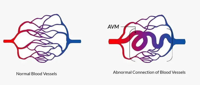
An irregular link between an artery and vein that avoids the capillary bed is called an arteriovenous fistula.
vascular malformations: Deformity exhibiting typical endothelium.
Angioma/hemangioma: Deformity characterized by endothelial hyperplasia.
Etiology
Congenital: categorized into malformations (AVMs) and haemangiomas (strawberry naevi, for example). The latter is separated into high flow and low flow types (such as pulmonary or hepatic AVM;
genetic: AVMs and haemangiomas are linked to a variety of genetic disorders, including hereditary hemorrhagic telangiectasia (Osler-Weber-Rendu syndrome) and Klippel–Trenaunay, Kasabach–Merritt, Sturge–Weber, and von Hippel–Lindau.
Aorto-venocaval fistula, glomus tumor, hypernephroma, sarcomas, infections, inflammations, or iatrogenic conditions (e.g., Brescia-Cimino fistula for hemodialysate or portocaval shunt in portal hypertension) are examples of acquired conditions.
Epidemiology
The others are less prevalent than cutaneous haemangiomas.
History
Depending on the location and extent of the AVM, symptoms might manifest differently and may result from either local or systemic effects (see Complications).
Congenital cutaneous haemangiomas frequently become apparent at birth or shortly thereafter.
Typically, malformations enlarge with aging, puberty, or pregnancy.
Internal organs may not exhibit symptoms until issues arise.
Other symptoms could be discomfort or swelling in the limbs, or varicose veins.
Examination
Cutaneous haemangiomas, also known as Campbell de Morgan spots, are often firm, crimson in color, and blood-filled when compressed.
An overlaying bruit or palpable thrill, possibly accompanied by decreased distal pulses and increased pulse pressure, may indicate the presence of internal AVMs.
Investigational studies
AVM imaging: Depending on where the lesion is located. Among the modalities employed are invasive angiography, CT or MRI scanning, and duplex scanning.
SPECT scan: Radiolabelled microspheres that are too big to go through capillaries are injected into an artery to quantify AV shunting. Using a gamma camera, those going through AVMs are trapped in the lungs and measured.
Management
Conservative: By the end of the first year of life, cutaneous haemangiomas typically spontaneously recede. Internal organ AVMs can be watched and may not require treatment.
Interventional radiology: Metal coils, tissue adhesive, or particles can be used to embolize internal AVMs or fistulae.
Surgery: Excision (following pre-operative embolization) is often challenging, although it is a possibility for small and easily accessible AVMs.
Useful for tiny AVMs, stereotactic radiosurgery may take years to fully manifest.
Complications
Cutaneous: bleeding, ulcers, and cosmetic deformity.
Organ-specific: For example, lung AVMs might result in hemoptysis or paradoxical embolism; brain AVMs can induce localized neurological impairments, convulsions, or stroke.
Peripheral tissue ischemia is considered distal.
Systemic: When there are significant AVMs, high-output heart failure may occur.
Prognosis
Depending on the aetiology and site. 90% of haemangiomas regress over a period of 5 to 10 years, while cerebral AVMs carry a 1-4 percent yearly risk of hemorrhage.
Introduction
An irregular link between an artery and vein that avoids the capillary bed is called an arteriovenous fistula.
vascular malformations: Deformity exhibiting typical endothelium.
Angioma/hemangioma: Deformity characterized by endothelial hyperplasia.
Etiology
Congenital: categorized into malformations (AVMs) and haemangiomas (strawberry naevi, for example). The latter is separated into high flow and low flow types (such as pulmonary or hepatic AVM;
genetic: AVMs and haemangiomas are linked to a variety of genetic disorders, including hereditary hemorrhagic telangiectasia (Osler-Weber-Rendu syndrome) and Klippel–Trenaunay, Kasabach–Merritt, Sturge–Weber, and von Hippel–Lindau.
Aorto-venocaval fistula, glomus tumor, hypernephroma, sarcomas, infections, inflammations, or iatrogenic conditions (e.g., Brescia-Cimino fistula for hemodialysate or portocaval shunt in portal hypertension) are examples of acquired conditions.
Epidemiology
The others are less prevalent than cutaneous haemangiomas.
History
Depending on the location and extent of the AVM, symptoms might manifest differently and may result from either local or systemic effects (see Complications).
Congenital cutaneous haemangiomas frequently become apparent at birth or shortly thereafter.
Typically, malformations enlarge with aging, puberty, or pregnancy.
Internal organs may not exhibit symptoms until issues arise.
Other symptoms could be discomfort or swelling in the limbs, or varicose veins.
Examination
Cutaneous haemangiomas, also known as Campbell de Morgan spots, are often firm, crimson in color, and blood-filled when compressed.
An overlaying bruit or palpable thrill, possibly accompanied by decreased distal pulses and increased pulse pressure, may indicate the presence of internal AVMs.
Investigational studies
AVM imaging: Depending on where the lesion is located. Among the modalities employed are invasive angiography, CT or MRI scanning, and duplex scanning.
SPECT scan: Radiolabelled microspheres that are too big to go through capillaries are injected into an artery to quantify AV shunting. Using a gamma camera, those going through AVMs are trapped in the lungs and measured.
Management
Conservative: By the end of the first year of life, cutaneous haemangiomas typically spontaneously recede. Internal organ AVMs can be watched and may not require treatment.
Interventional radiology: Metal coils, tissue adhesive, or particles can be used to embolize internal AVMs or fistulae.
Surgery: Excision (following pre-operative embolization) is often challenging, although it is a possibility for small and easily accessible AVMs.
Useful for tiny AVMs, stereotactic radiosurgery may take years to fully manifest.
Complications
Cutaneous: bleeding, ulcers, and cosmetic deformity.
Organ-specific: For example, lung AVMs might result in hemoptysis or paradoxical embolism; brain AVMs can induce localized neurological impairments, convulsions, or stroke.
Peripheral tissue ischemia is considered distal.
Systemic: When there are significant AVMs, high-output heart failure may occur.
Prognosis
Depending on the aetiology and site. 90% of haemangiomas regress over a period of 5 to 10 years, while cerebral AVMs carry a 1-4 percent yearly risk of hemorrhage.
0 Comments
