- Published on
Surgery -Below The knee Amputation
Indications
gangrene, ischemia, or infarction: severe trauma, burns, or lower limb ischaemia, either acute or chronic.
Some tumors are malignant (e.g., osteosarcoma and malignant melanoma).
Severe infection: necrotizing fasciitis or gas gangrene (Clostridium perfringens).
Unmanageable ulcers or excruciatingly paralyzed limbs are uncommon.
Anatomy
Below the knee: Robinson's skew flap and Burgess long posterior flap are the two transtibial amputation procedures.
Ankle level: Rarely used since prosthesis attachment is challenging.
Midfoot: Chopart's disarticulation of the talonavicular and calcaneocuboid joints or Lisfranc's involving the disarticulation between the tarsal and metatarsal bones.
Ray: This involves cutting through the metatarsal bone to remove a toe.
Toe: Because cutting through a joint exposes avascular cartilage that is poorly mending, division occurs through the proximal phalanx.
Investigations
Pre-operative care should ideally involve a comprehensive evaluation by professionals in surgery, anesthesia, prosthetics, physiotherapy, psychology, and other fields. Determining the degree of amputation in light of the patient's characteristics and the severity of the condition (e.g. rehabilitation chances). If a patient has diabetes, a sliding insulin scale, suitable blood tests and cross-matching, and, if necessary, urine catheterization.
Following surgery: Early physiotherapy, prosthesis fitting, or walking aids (such as a pneumatic postamputation mobility device) are used in the rehabilitation process.
Procedure
Access: Skin flaps are marked on the skin before the incision, either asymmetrical anteromedial and posterolateral flaps or a lengthier posterior flap (Burgess). Tibial transition occurs 10–12 cm below tibial tuberosity or 14 cm below the knee joint.
Ligation of muscle and vessels: During a skin incision, the anterior and peroneal compartment muscles are separated by diathermy, and the long saphenous vein is ligated. Veins and arteries are tied off, and the tibial nerve is neatly separated under light traction when the concomitant vasa nervorum dilates.
Amputation of the bone: After the periosteum is removed, the fibula is split 2 cm proximally.
In addition, the tibia is separated and stripped, and the ends of the bones are filed smooth.
Closure: A cylindrical stump is formed by the posterior flap covering the severed tibia with a portion of the gastrocnemius muscle. Sutures are used to seal the skin after hemostasis is reached.
You might leave a drain in place.
Complications
Early: Bony spurs, psychological issues, stump length that is too long or short, stump hemorrhage, DVT, flap ischaemia, and pain.
Late: creation of neuromas, erosion of bone through skin, ischaemia, osteomyelitis, ulceration, and "phantom" limb pain (which is lessened by effective analgesia after surgery).
Prognosis
Amputations are most frequently performed on patients who also have significant atherosclerotic disease, and only 30% of these patients survive after five years.
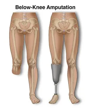
Refer to Fig. 20 for a diagrammatic summary of general amputations.
Indications
gangrene, ischemia, or infarction: severe trauma, burns, or lower limb ischaemia, either acute or chronic.
Some tumors are malignant (e.g., osteosarcoma and malignant melanoma).
Severe infection: necrotizing fasciitis or gas gangrene (Clostridium perfringens).
Unmanageable ulcers or excruciatingly paralyzed limbs are uncommon.
Anatomy
Below the knee: Robinson's skew flap and Burgess long posterior flap are the two transtibial amputation procedures.
Ankle level: Rarely used since prosthesis attachment is challenging.
Midfoot: Chopart's disarticulation of the talonavicular and calcaneocuboid joints or Lisfranc's involving the disarticulation between the tarsal and metatarsal bones.
Ray: This involves cutting through the metatarsal bone to remove a toe.
Toe: Because cutting through a joint exposes avascular cartilage that is poorly mending, division occurs through the proximal phalanx.
Investigations
Pre-operative care should ideally involve a comprehensive evaluation by professionals in surgery, anesthesia, prosthetics, physiotherapy, psychology, and other fields. Determining the degree of amputation in light of the patient's characteristics and the severity of the condition (e.g. rehabilitation chances). If a patient has diabetes, a sliding insulin scale, suitable blood tests and cross-matching, and, if necessary, urine catheterization.
Following surgery: Early physiotherapy, prosthesis fitting, or walking aids (such as a pneumatic postamputation mobility device) are used in the rehabilitation process.
Procedure
Access: Skin flaps are marked on the skin before the incision, either asymmetrical anteromedial and posterolateral flaps or a lengthier posterior flap (Burgess). Tibial transition occurs 10–12 cm below tibial tuberosity or 14 cm below the knee joint.
Ligation of muscle and vessels: During a skin incision, the anterior and peroneal compartment muscles are separated by diathermy, and the long saphenous vein is ligated. Veins and arteries are tied off, and the tibial nerve is neatly separated under light traction when the concomitant vasa nervorum dilates.
Amputation of the bone: After the periosteum is removed, the fibula is split 2 cm proximally.
In addition, the tibia is separated and stripped, and the ends of the bones are filed smooth.
Closure: A cylindrical stump is formed by the posterior flap covering the severed tibia with a portion of the gastrocnemius muscle. Sutures are used to seal the skin after hemostasis is reached.
You might leave a drain in place.
Complications
Early: Bony spurs, psychological issues, stump length that is too long or short, stump hemorrhage, DVT, flap ischaemia, and pain.
Late: creation of neuromas, erosion of bone through skin, ischaemia, osteomyelitis, ulceration, and "phantom" limb pain (which is lessened by effective analgesia after surgery).
Prognosis
Amputations are most frequently performed on patients who also have significant atherosclerotic disease, and only 30% of these patients survive after five years.
Refer to Fig. 20 for a diagrammatic summary of general amputations.
0 Comments
