- Published on
Surgery - Benign Breast Disease
Definition
breast diseases that are not cancerous, such as physiopathological abnormalities of the adipose, stromal, epithelial, or vascular components of the breast.
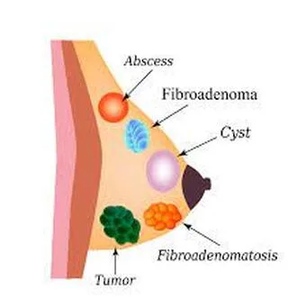
Fibroadenoma: Occurs when a breast lobule hyperplasia occurs and comprises both normal connective tissue and epithelial cells.
Fat necrosis: Amorphous material, inflammatory cells, including foreign body giant cells, and irregular and necrotic adipocytes can all resemble cancer.
A normal involution is aberrated in sclerosing adenosis.
Duct ectasia: This condition is brought about by central ducts becoming dilated with secretions; if leaking into periductal tissue happens, it can result in periductal mastitis, an inflammatory response.
Origin
Under endocrine regulation, breast tissue changes in a variety of ways. Trauma is the secondary cause of fat necrosis. Benign conditions are mapped using the ANDI (aberrations of normal development and involution) classification system based on the degree of abnormality as well as the pathophysiology.
Risk Factors
possibly less common in people using birth control pills. One risk factor for periductal masttitis is smoking.
The study of epidemiology
Only 10–20% of instances are thought to proceed to a histopathological diagnosis. As many as 60% of women have diffuse fibrocystic alterations, and 70% of them have mastalgia.
Breast cysts are more prevalent in people 40 to 50 years old, and fibroadenomas are more common in people 15 to 25 years old. Unless a woman is on hormone replacement therapy (HRT), these conditions often go away after menopause.
History
Past history of lump or enlargement, breast pain or discomfort (cyclical or non-cyclical mastalgia).
Breast discharge: cancer should be suspected if it is bloodstained. It is important to determine risk factors for breast cancer, such as menstruation history, family history, pregnancy history, usage of OCP, and hormone replacement treatment.
Examination
Breast nodules, focal or widespread.
Typically, fibroadenomas are smooth, well-circumscribed, and movable lumps that have a diameter of 1-2 cm in breast mice.
Nipple discharge that is yellow or green (duct ectasia).
There are no signs of malignancy, such as dimpling, peau d'orange skin changes, or swollen lymph nodes in the arms.
Investigational studies
Typically carried out in conjunction with triple assessment:
1. A clinical assessment.
2. Imaging: USS in younger patients (<35 years old) or mammography (craniocaudal and oblique mediolateral views, spot compression and magnification views). Less frequently, benign lumps will become calcified (microcalcifications strongly signal malignancy). MRI examinations are also beneficial.
3. Cytology/Histology: By trucut or excision biopsy, or by FNA (fine-needle aspiration) cytology.
Conservative management includes treating the symptoms, such as analgesia and mastalgia with evening primrose oil, which is a rich source of gammalinoleic acid. Tips for eating a lower-fat diet and using supportive bras.Danazol is applied as a backup therapy. 17-a-ethinyl testosterone inhibits the production of ovarian steroids, reduces gonadotropin release, and stops the LH surge.
If fibroadenomas are significant or the patient requests it, they can be removed or treated cautiously.
Simple cysts should totally vanish after aspiration and don't require aspiration unless clinically required. In the absence of that, treat it like a breast lump.
Surgery: This involves removing or excising the breast lump through biopsy; if there is any reason to suspect that the lump is not benign, a broad local excision should be carried out. The treatment for intraductal papillomas is microdochectomy. In duct ectasia, the Hadfields (or Adairs) procedure excises the central ducts.
Complications
Pain reappearance.
The prognosis is good, although recurrence is frequent. Fibroadenomas: women with simple FA do not have an elevated risk of cancer, nor is there a higher family history of breast cancer.
Definition
breast diseases that are not cancerous, such as physiopathological abnormalities of the adipose, stromal, epithelial, or vascular components of the breast.
Fibroadenoma: Occurs when a breast lobule hyperplasia occurs and comprises both normal connective tissue and epithelial cells.
Fat necrosis: Amorphous material, inflammatory cells, including foreign body giant cells, and irregular and necrotic adipocytes can all resemble cancer.
A normal involution is aberrated in sclerosing adenosis.
Duct ectasia: This condition is brought about by central ducts becoming dilated with secretions; if leaking into periductal tissue happens, it can result in periductal mastitis, an inflammatory response.
Origin
Under endocrine regulation, breast tissue changes in a variety of ways. Trauma is the secondary cause of fat necrosis. Benign conditions are mapped using the ANDI (aberrations of normal development and involution) classification system based on the degree of abnormality as well as the pathophysiology.
Risk Factors
possibly less common in people using birth control pills. One risk factor for periductal masttitis is smoking.
The study of epidemiology
Only 10–20% of instances are thought to proceed to a histopathological diagnosis. As many as 60% of women have diffuse fibrocystic alterations, and 70% of them have mastalgia.
Breast cysts are more prevalent in people 40 to 50 years old, and fibroadenomas are more common in people 15 to 25 years old. Unless a woman is on hormone replacement therapy (HRT), these conditions often go away after menopause.
History
Past history of lump or enlargement, breast pain or discomfort (cyclical or non-cyclical mastalgia).
Breast discharge: cancer should be suspected if it is bloodstained. It is important to determine risk factors for breast cancer, such as menstruation history, family history, pregnancy history, usage of OCP, and hormone replacement treatment.
Examination
Breast nodules, focal or widespread.
Typically, fibroadenomas are smooth, well-circumscribed, and movable lumps that have a diameter of 1-2 cm in breast mice.
Nipple discharge that is yellow or green (duct ectasia).
There are no signs of malignancy, such as dimpling, peau d'orange skin changes, or swollen lymph nodes in the arms.
Investigational studies
Typically carried out in conjunction with triple assessment:
1. A clinical assessment.
2. Imaging: USS in younger patients (<35 years old) or mammography (craniocaudal and oblique mediolateral views, spot compression and magnification views). Less frequently, benign lumps will become calcified (microcalcifications strongly signal malignancy). MRI examinations are also beneficial.
3. Cytology/Histology: By trucut or excision biopsy, or by FNA (fine-needle aspiration) cytology.
Conservative management includes treating the symptoms, such as analgesia and mastalgia with evening primrose oil, which is a rich source of gammalinoleic acid. Tips for eating a lower-fat diet and using supportive bras.Danazol is applied as a backup therapy. 17-a-ethinyl testosterone inhibits the production of ovarian steroids, reduces gonadotropin release, and stops the LH surge.
If fibroadenomas are significant or the patient requests it, they can be removed or treated cautiously.
Simple cysts should totally vanish after aspiration and don't require aspiration unless clinically required. In the absence of that, treat it like a breast lump.
Surgery: This involves removing or excising the breast lump through biopsy; if there is any reason to suspect that the lump is not benign, a broad local excision should be carried out. The treatment for intraductal papillomas is microdochectomy. In duct ectasia, the Hadfields (or Adairs) procedure excises the central ducts.
Complications
Pain reappearance.
The prognosis is good, although recurrence is frequent. Fibroadenomas: women with simple FA do not have an elevated risk of cancer, nor is there a higher family history of breast cancer.
0 Comments
