- Published on
Surgery - Breast Cancer
Overview
Cancer that originates in the breast tissue.
Origin
a mix of environmental and genetic variables.
Genetics: 5–10% of instances are due to genetic factors, but the majority are polygenic risk. 2% of cases contain BRCA-1 (17q) and BRCA-2 (13q) gene mutations, with carriers having a lifetime risk of up to 87%. Li-Fraumeni syndrome (TP53), Cowden's syndrome (PTEN), Peutz-Jeghers syndrome (STK11/LKB1), ataxia-telagiectasia (ATM), and Muir-Torre syndrome (MSH2/MLH1) are examples of rare hereditary breast cancer syndromes.
Risk factors include age, nulliparity, early menarche, late menopause, menopausal hormone replacement medication, obesity, alcohol consumption, and extended exposure to female sex hormones, especially oestrogen.
Epidemiology is the second most common cause of cancer-related deaths in women worldwide, after lung cancer in the United States. In the UK, the lifetime risk is 1 in 9. peak incidence in people aged 40 to 70. Just 1% of all breast cancers in men are rare.
History
perhaps found by screening.
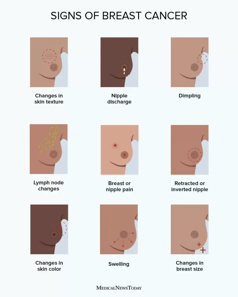
Primary symptoms include breast lumps, which are typically painless, breast form alterations, and nipple discharge.
Axillary lump, bone pain, weight loss, and paraneoplastic disorders (such as cerebellar syndrome) are signs of secondary spread.
Examination
When the patient is supine and upright, the breasts should be examined for asymmetry, peau d'orange (oedema), dimpling or tethering, nipple scaling or inversion, or, in more severe situations, ulceration.
Use a clockwise radial approach for palpating hard, irregular, or fixed lumps.
Check for hepatomegaly, chest anomalies, palpable axillary lymph nodes, supraclavicular lymph nodes, and bone discomfort.
Investigations
Triple assessment: A standardized procedure that includes a clinical evaluation, imaging (MRI, CT, or mammography), and tissue diagnosis (cytology or biopsy) to investigate a breast lump.
Mammography (Fig. 1): A valuable screening tool for women over 35. In the UK, screening doesn't start until beyond age 50. The craniocaudal and mediolateral oblique views are typical. Spiculated lesions and branching or linear microcalcifications are characteristics of malignancy.
Ultrasound: To distinguish between dangerous solid lesions and benign cystic lesions. More beneficial for women under 35.
Fine-needle aspiration: Less invasive, enables cyst drainage and cytology of individual breast masses.
Core biopsy: Allows for histological diagnosis; can be image guided.
Sentinel lymph node biopsy: After injecting radioactive tracer or blue dye in the vicinity of the breast lesion, a nuclear scan locates the sentinel node, which is then biopsied to look for spread.
CT (chest, abdomen, pelvis), PET, or bone scans for metastases are the staging options.
Blood: CA-15-3 tumor marker, bone profile, LFT, FBC, U&Es, and Ca2₠.
Histopathology:
In situ cancer:non-invasive ductal or lobular carcinoma in situ (DCIS, LCIS) that preserves the basement membrane.
Invasive: Ductal carcinoma accounts for 75% of all breast cancer cases.
Other: tubular, mucinous, medullary, cruriform, papillary, lobular (10–15%, Indian filing arrangement of cells), and Paget's disease of the nipple (ductal carcinoma in situ infiltrating the nipple).
Phlloides: Fibroepithelial tumours that can be benign or malignant.
Molecular prognostic factors: Oestrogen and progesterone receptors (ER, PR) and HER-2
expression (20–30% of cancers) are valuable prognostic indicators and guide treatment.
Flow cytometry measures DNA content (ploidy) and S-phase fraction (cell proliferation
rate).
Grading: The Nottingham modification of the Bloom and Richardson grading system is a
prognostic indicator. Three features assessed are tubule formation, nuclear size/
pleomorphism and number of mitoses. Scores are used to generate Grades 1 (well
differentiated) to 3 (poorly differentiated).
Staging: The UICC TNM-staging system.
Tumour size (T): T1: <2 cm; T2: 2–5 cm; T3: >5 cm; T4: any size with chest wall or skin
extension.
Nodes (N): N1: mobile ipsilateral axillary; N2: fixed ipsilateral axillary; N3: ipsilateral internal
mammary nodes.
Metastases (M): M0: no distant metastases; M1: distant metastases.
Supervisory
Breast care nurses, radiologists, oncologists, and breast surgeons are all part of the multidisciplinary management team. The size, location, type, stage, and consideration of each patient's unique wishes all play a role in the decision to remove the cancer surgically.
Breast-conserving surgery: Segmental mastectomy or wide local excision (if the patient is willing to receive radiation therapy and the single cancer is less than 5 cm). Radiological wire localization can be required for smaller lesions.
Modified radical mastectomy: dissection of the axillary lymph nodes and total mastectomy.
Axillary surgery is required for node staging and can vary from level III clearance (lymph nodes up to and above the pectoralis minor muscle) to sentinel node biopsy, which removes three nodes on average.
Breast reconstruction: Usually done after the surgery, but sometimes it happens right away after surgical excision. Techniques include transverse rectus abdominis myocutaneous flaps, latissimus dorsi flaps, and breast prosthesis.
Radiotherapy: After breast-conserving surgery, external beam radiation is sometimes used as neoadjuvant therapy and to palliate advanced tumors.
Chemotherapy: Adjuvant, palliative, and neoadjuvant settings are possible for treatment. Visceral involvement, oestrogen receptor-negative tumors, fast-progressing illness, or failure of hormonal therapy are more common in premenopausal women.
Combination therapy regimens, such as those involving cyclophosphamide, methotrexate, and 5-fluorouracil (CMF), are customized for each patient.
The primary first-line treatment for oestrogen receptor-positive tumors is hormonal therapy, which includes selective oestrogen receptor modulators like tamoxifen. Additional options include ovarian ablation using LHRH analogs, such as goserelin, aromatase inhibitors in postmenopausal women, such as letrozole or anastrozole, and selective oestrogen receptor downregulators, such as progestins and fullvestrant.
Biological therapy: In nodal and HER-2-positive cancer, trastuzumab (Herceptin), a monoclonal antibody that targets the HER-2 receptor (a cell growth promoter), is used in conjunction with chemotherapy and has been demonstrated to increase overall and disease-free survival.
Complications
significant physical or psychological damage following surgery or diagnosis.
Hypercalcemia, spinal cord compression, bone pain, and problems with the abdomen, lungs, or brain can all result from metastases.
Venous thrombosis and endometrial cancer are caused by tamoxifen. Osteoporosis, joint/muscle pain, aromatase inhibitors. The cardiotoxicity of herceptin.
Following surgery, there may be a local recurrence, wound infection, hemorrhage, lymphoedema, shoulder pain, sensory loss (the intercostobrachial nerve is frequently sacrificed, resulting in numbness in the inner, upper arm).
Fatigue, skin alterations, and lymphoedema are side effects of radiation therapy.
Prognosis varies according on stage, grade, and kind. Total 5-year survival is 100% if the illness is limited to the breast, 50%–90% if it is node-positive, and 20% if it has spread to distant organs.tumors of the fibroepithelium that may be benign or cancerous.
& HER-2 expression (20–30% of malignancies) and oestrogen and progesterone receptors (ER, PR) are important prognostic markers that help determine treatment.
DNA content (ploidy) and S-phase fraction (cell proliferation rate) are measured by flow cytometry.
Grading: A prognostic indicator is the Nottingham version of the Bloom and Richardson grading system. Nuclear size/pleomorphism, quantity of mitoses, and tubule development are the three characteristics that are evaluated. Grades 1 (highly differentiated) through 3 (poorly differentiated) are created using scores.
UICC TNM-staging system is used for staging.
Tumor size (T): <2 cm for T1, 2–5 cm for T2, >5 cm for T3, and any size with skin or chest wall expansion for T4.
Nodes (N): ipsilateral internal mammary nodes (N3), mobile ipsilateral axillary nodes (N1), and stationary ipsilateral axillary nodes (N2).
Metastases (M): M0 denotes no distant metastases, while M1 denotes distant metastases.
Overview
Cancer that originates in the breast tissue.
Origin
a mix of environmental and genetic variables.
Genetics: 5–10% of instances are due to genetic factors, but the majority are polygenic risk. 2% of cases contain BRCA-1 (17q) and BRCA-2 (13q) gene mutations, with carriers having a lifetime risk of up to 87%. Li-Fraumeni syndrome (TP53), Cowden's syndrome (PTEN), Peutz-Jeghers syndrome (STK11/LKB1), ataxia-telagiectasia (ATM), and Muir-Torre syndrome (MSH2/MLH1) are examples of rare hereditary breast cancer syndromes.
Risk factors include age, nulliparity, early menarche, late menopause, menopausal hormone replacement medication, obesity, alcohol consumption, and extended exposure to female sex hormones, especially oestrogen.
Epidemiology is the second most common cause of cancer-related deaths in women worldwide, after lung cancer in the United States. In the UK, the lifetime risk is 1 in 9. peak incidence in people aged 40 to 70. Just 1% of all breast cancers in men are rare.
History
perhaps found by screening.
Primary symptoms include breast lumps, which are typically painless, breast form alterations, and nipple discharge.
Axillary lump, bone pain, weight loss, and paraneoplastic disorders (such as cerebellar syndrome) are signs of secondary spread.
Examination
When the patient is supine and upright, the breasts should be examined for asymmetry, peau d'orange (oedema), dimpling or tethering, nipple scaling or inversion, or, in more severe situations, ulceration.
Use a clockwise radial approach for palpating hard, irregular, or fixed lumps.
Check for hepatomegaly, chest anomalies, palpable axillary lymph nodes, supraclavicular lymph nodes, and bone discomfort.
Investigations
Triple assessment: A standardized procedure that includes a clinical evaluation, imaging (MRI, CT, or mammography), and tissue diagnosis (cytology or biopsy) to investigate a breast lump.
Mammography (Fig. 1): A valuable screening tool for women over 35. In the UK, screening doesn't start until beyond age 50. The craniocaudal and mediolateral oblique views are typical. Spiculated lesions and branching or linear microcalcifications are characteristics of malignancy.
Ultrasound: To distinguish between dangerous solid lesions and benign cystic lesions. More beneficial for women under 35.
Fine-needle aspiration: Less invasive, enables cyst drainage and cytology of individual breast masses.
Core biopsy: Allows for histological diagnosis; can be image guided.
Sentinel lymph node biopsy: After injecting radioactive tracer or blue dye in the vicinity of the breast lesion, a nuclear scan locates the sentinel node, which is then biopsied to look for spread.
CT (chest, abdomen, pelvis), PET, or bone scans for metastases are the staging options.
Blood: CA-15-3 tumor marker, bone profile, LFT, FBC, U&Es, and Ca2₠.
Histopathology:
In situ cancer:non-invasive ductal or lobular carcinoma in situ (DCIS, LCIS) that preserves the basement membrane.
Invasive: Ductal carcinoma accounts for 75% of all breast cancer cases.
Other: tubular, mucinous, medullary, cruriform, papillary, lobular (10–15%, Indian filing arrangement of cells), and Paget's disease of the nipple (ductal carcinoma in situ infiltrating the nipple).
Phlloides: Fibroepithelial tumours that can be benign or malignant.
Molecular prognostic factors: Oestrogen and progesterone receptors (ER, PR) and HER-2
expression (20–30% of cancers) are valuable prognostic indicators and guide treatment.
Flow cytometry measures DNA content (ploidy) and S-phase fraction (cell proliferation
rate).
Grading: The Nottingham modification of the Bloom and Richardson grading system is a
prognostic indicator. Three features assessed are tubule formation, nuclear size/
pleomorphism and number of mitoses. Scores are used to generate Grades 1 (well
differentiated) to 3 (poorly differentiated).
Staging: The UICC TNM-staging system.
Tumour size (T): T1: <2 cm; T2: 2–5 cm; T3: >5 cm; T4: any size with chest wall or skin
extension.
Nodes (N): N1: mobile ipsilateral axillary; N2: fixed ipsilateral axillary; N3: ipsilateral internal
mammary nodes.
Metastases (M): M0: no distant metastases; M1: distant metastases.
Supervisory
Breast care nurses, radiologists, oncologists, and breast surgeons are all part of the multidisciplinary management team. The size, location, type, stage, and consideration of each patient's unique wishes all play a role in the decision to remove the cancer surgically.
Breast-conserving surgery: Segmental mastectomy or wide local excision (if the patient is willing to receive radiation therapy and the single cancer is less than 5 cm). Radiological wire localization can be required for smaller lesions.
Modified radical mastectomy: dissection of the axillary lymph nodes and total mastectomy.
Axillary surgery is required for node staging and can vary from level III clearance (lymph nodes up to and above the pectoralis minor muscle) to sentinel node biopsy, which removes three nodes on average.
Breast reconstruction: Usually done after the surgery, but sometimes it happens right away after surgical excision. Techniques include transverse rectus abdominis myocutaneous flaps, latissimus dorsi flaps, and breast prosthesis.
Radiotherapy: After breast-conserving surgery, external beam radiation is sometimes used as neoadjuvant therapy and to palliate advanced tumors.
Chemotherapy: Adjuvant, palliative, and neoadjuvant settings are possible for treatment. Visceral involvement, oestrogen receptor-negative tumors, fast-progressing illness, or failure of hormonal therapy are more common in premenopausal women.
Combination therapy regimens, such as those involving cyclophosphamide, methotrexate, and 5-fluorouracil (CMF), are customized for each patient.
The primary first-line treatment for oestrogen receptor-positive tumors is hormonal therapy, which includes selective oestrogen receptor modulators like tamoxifen. Additional options include ovarian ablation using LHRH analogs, such as goserelin, aromatase inhibitors in postmenopausal women, such as letrozole or anastrozole, and selective oestrogen receptor downregulators, such as progestins and fullvestrant.
Biological therapy: In nodal and HER-2-positive cancer, trastuzumab (Herceptin), a monoclonal antibody that targets the HER-2 receptor (a cell growth promoter), is used in conjunction with chemotherapy and has been demonstrated to increase overall and disease-free survival.
Complications
significant physical or psychological damage following surgery or diagnosis.
Hypercalcemia, spinal cord compression, bone pain, and problems with the abdomen, lungs, or brain can all result from metastases.
Venous thrombosis and endometrial cancer are caused by tamoxifen. Osteoporosis, joint/muscle pain, aromatase inhibitors. The cardiotoxicity of herceptin.
Following surgery, there may be a local recurrence, wound infection, hemorrhage, lymphoedema, shoulder pain, sensory loss (the intercostobrachial nerve is frequently sacrificed, resulting in numbness in the inner, upper arm).
Fatigue, skin alterations, and lymphoedema are side effects of radiation therapy.
Prognosis varies according on stage, grade, and kind. Total 5-year survival is 100% if the illness is limited to the breast, 50%–90% if it is node-positive, and 20% if it has spread to distant organs.tumors of the fibroepithelium that may be benign or cancerous.
& HER-2 expression (20–30% of malignancies) and oestrogen and progesterone receptors (ER, PR) are important prognostic markers that help determine treatment.
DNA content (ploidy) and S-phase fraction (cell proliferation rate) are measured by flow cytometry.
Grading: A prognostic indicator is the Nottingham version of the Bloom and Richardson grading system. Nuclear size/pleomorphism, quantity of mitoses, and tubule development are the three characteristics that are evaluated. Grades 1 (highly differentiated) through 3 (poorly differentiated) are created using scores.
UICC TNM-staging system is used for staging.
Tumor size (T): <2 cm for T1, 2–5 cm for T2, >5 cm for T3, and any size with skin or chest wall expansion for T4.
Nodes (N): ipsilateral internal mammary nodes (N3), mobile ipsilateral axillary nodes (N1), and stationary ipsilateral axillary nodes (N2).
Metastases (M): M0 denotes no distant metastases, while M1 denotes distant metastases.
0 Comments
