- Published on
Surgery - Cardiac Transplantation
Indications
99% of cases are of ischemic heart disease and idiopathic cardiomyopathy.
Myocarditis (1%), valvular heart disease, and congenital heart disease.
Qualifications for recipients:. end-stage heart disease with less than a year to live.
lack of renal or hepatic dysfunction.
lack of a tumor or active systemic infection.
psychosocial stability and the capacity to adhere to immunosuppressive treatment and aftercare.
Anatomy
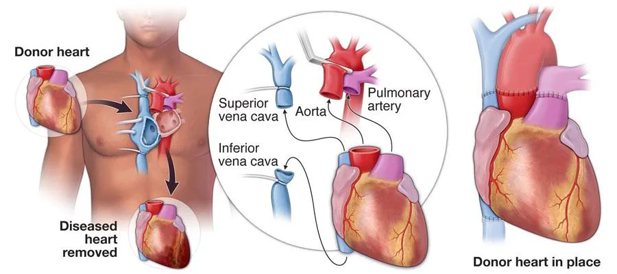
Orthotopic transplantation: The majority of the recipient's heart is replaced by the donor heart. The ventricles are removed from the latter, leaving the large vessels—the right and left atriums—in place.
The left atria, followed by the right atria, and finally the large vessels, are anastomosed to the donor heart.
As an alternative, bicaval anastamosis (which reduces atrial and valvular issues) can be carried out.
Heterotopic transplantation involves positioning the donor heart parallel to the recipient's heart without removing the recipient's heart. less frequently carried out. The recipient's heart is aided by the donor heart. is feasible when pulmonary hypertension is present.
Investigations
Prior to surgery: comprehensive interdisciplinary workup that includes tissue typing, panel-reactive antibodies, blood testing, blood grouping, and infection screening. Anaesthetic evaluation.
Imaging: CXR, echo, and coronary angiography. testing for pulmonary function. Evaluation of maximal venous oxygen consumption (MVO2), a measure of the severity of heart failure, and pulmonary vascular resistance.
Donors: Heart beating and brain stem death requirements met in an ABO compatible manner. must not have any heart pathologies. A team of experts performs donor surgery. The heart can only withstand brief preservation periods—up to six hours.
After surgery, patients are first closely monitored in intensive care.
Immunosuppressive medication is administered, such as azathioprine or cyclosporin. Biopsies of the endomyocardium are performed to evaluate for graft rejection.
Procedure (Orthotopic )
Entry: Sternotomy median.
Cardiopulmonary bypass: Used during cardiac surgery to oxygenate and circulate blood.
Removal of recipient organ: A mid-atrial incision is performed across the recipient's left and right atria. Just prior to the left and right pulmonary arteries splitting off, the ascending aorta and the pulmonary artery split. The heart of the recipient is extracted.
Preparing a donor organ: The heart is meticulously ready for implantation.
Recipient transplantation (inflow anastomosis): Running sutures are used to anastomose the left atrium to the recipient's remaining left atria. To keep the donor heart cool, topical cooling with frozen saline slush is given externally.
After that, the recipient's remaining right atrium is anastomosed to the right atrium.
reestablishing outflow anastomosis and circulation: As the pulmonary artery and aorta are being anastomosed, systemic rewarming is initiated. Either spontaneously or with cardioversion, sinus rhythm recovers. The bypass circuit is progressively turned off for the patient, enabling the heart to fill and start the cardiac ejection process again. All cannulas are withdrawn once cardiac output is deemed sufficient.
Closure: Two thoracostomy drains are placed in the chest cavity and pericardium, and temporary pacing wires are implanted into the atrium and/or ventricles. The skin is closed and the sternum is sealed with broken steel wires.
Complications
Early symptoms include hemorrhage, infection, organ failure, severe rejection, and arrhythmias.
Late: Hypertension, chronic rejection, infection, valve regurgitation, accelerated coronary artery disease, and post-transplantation malignancy.
Prognosis
3-5% is the operational mortality rate. 90% of people survive one year, and 78% survive five.
Indications
99% of cases are of ischemic heart disease and idiopathic cardiomyopathy.
Myocarditis (1%), valvular heart disease, and congenital heart disease.
Qualifications for recipients:. end-stage heart disease with less than a year to live.
lack of renal or hepatic dysfunction.
lack of a tumor or active systemic infection.
psychosocial stability and the capacity to adhere to immunosuppressive treatment and aftercare.
Anatomy
Orthotopic transplantation: The majority of the recipient's heart is replaced by the donor heart. The ventricles are removed from the latter, leaving the large vessels—the right and left atriums—in place.
The left atria, followed by the right atria, and finally the large vessels, are anastomosed to the donor heart.
As an alternative, bicaval anastamosis (which reduces atrial and valvular issues) can be carried out.
Heterotopic transplantation involves positioning the donor heart parallel to the recipient's heart without removing the recipient's heart. less frequently carried out. The recipient's heart is aided by the donor heart. is feasible when pulmonary hypertension is present.
Investigations
Prior to surgery: comprehensive interdisciplinary workup that includes tissue typing, panel-reactive antibodies, blood testing, blood grouping, and infection screening. Anaesthetic evaluation.
Imaging: CXR, echo, and coronary angiography. testing for pulmonary function. Evaluation of maximal venous oxygen consumption (MVO2), a measure of the severity of heart failure, and pulmonary vascular resistance.
Donors: Heart beating and brain stem death requirements met in an ABO compatible manner. must not have any heart pathologies. A team of experts performs donor surgery. The heart can only withstand brief preservation periods—up to six hours.
After surgery, patients are first closely monitored in intensive care.
Immunosuppressive medication is administered, such as azathioprine or cyclosporin. Biopsies of the endomyocardium are performed to evaluate for graft rejection.
Procedure (Orthotopic )
Entry: Sternotomy median.
Cardiopulmonary bypass: Used during cardiac surgery to oxygenate and circulate blood.
Removal of recipient organ: A mid-atrial incision is performed across the recipient's left and right atria. Just prior to the left and right pulmonary arteries splitting off, the ascending aorta and the pulmonary artery split. The heart of the recipient is extracted.
Preparing a donor organ: The heart is meticulously ready for implantation.
Recipient transplantation (inflow anastomosis): Running sutures are used to anastomose the left atrium to the recipient's remaining left atria. To keep the donor heart cool, topical cooling with frozen saline slush is given externally.
After that, the recipient's remaining right atrium is anastomosed to the right atrium.
reestablishing outflow anastomosis and circulation: As the pulmonary artery and aorta are being anastomosed, systemic rewarming is initiated. Either spontaneously or with cardioversion, sinus rhythm recovers. The bypass circuit is progressively turned off for the patient, enabling the heart to fill and start the cardiac ejection process again. All cannulas are withdrawn once cardiac output is deemed sufficient.
Closure: Two thoracostomy drains are placed in the chest cavity and pericardium, and temporary pacing wires are implanted into the atrium and/or ventricles. The skin is closed and the sternum is sealed with broken steel wires.
Complications
Early symptoms include hemorrhage, infection, organ failure, severe rejection, and arrhythmias.
Late: Hypertension, chronic rejection, infection, valve regurgitation, accelerated coronary artery disease, and post-transplantation malignancy.
Prognosis
3-5% is the operational mortality rate. 90% of people survive one year, and 78% survive five.
0 Comments
