- Published on
Surgery - Carotid Body Tumor
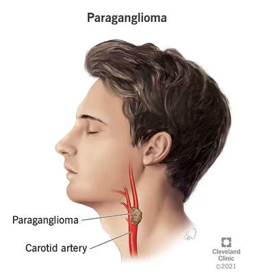
A chemodectoma is a tumor that develops from chemoreceptor cells at the carotid bifurcation.
Etiology
connected to changes in the mitochondrial succinate dehydrogenase complex subunits. With autosomal dominant inheritance, one-third are family (more likely to be bilateral and numerous).
Shamblin has classified:. Type I: Carotid bifurcation-derived little tumors that are easily dissected. Type II: More expansive and sticky, involving but not encasing vessels. Type III: Encircling the carotid bifurcation, large in size.
Risk factors linked to von Hippel-Lindau syndrome, MEN II, and NF1.
People who spend extended periods of time living at high elevations have a "incidence."
The study of epidemiology
uncommon. Present at any age; however, women are more likely than males to do so between the ages of 50 and 70.
History
Most frequently manifests as a gradually expanding lump in the neck. Symptoms include dysphagia, choking, and hoarseness may be brought on by pressure on surrounding cranial nerves.
Examination
A neck lump with transmitted pulse that is located in the carotid triangle of the neck. Horner syndrome with paresis or palsy of the cranial nerves VII, IX, X, and XI may be present.
Pathophysiology
Paragangliomas, which include glomus jugulare tumors, pheochromocytomas, and vagal body tumors, are tumors produced from neural crest tissue. One type of paraganglioma is the carotid body tumor.
Vascular tumor with sustentacular cells encircling clusters of zellballen of epithelioid "chief cells" Although they have the capacity to release catecholamines, the cells are often nonsecretory.
Investigations
Imaging: The link between the carotid bifurcation can be demonstrated by ultrasound and duplex scanning; angiography, which can be either CT or MR, verifies the typical splaying of the internal and external carotid arteries as well as the wine-glass-shaped tumor blush.
A CT or MRI scan is used to ascertain the tumor's extent. Metaiodobenzylguanidine (131I-MIBG) scintigraphy in functioning tumors.
Pharyngoscopy, both direct and indirect, can be used to evaluate pharyngeal invasion or involvement of cranial nerves.
Urine: VMA, metanephrine, and catecholamines.
Management
Surgery: Risk based on tumor size and extent. Due to their high vascularity, big tumors may be embolized prior to surgery (a controversial procedure), and preoperative blood should be cross-matched. Excision of the tumor surgically: Tumor size and place determine the incision. cautious jugular vein dissection and control, detection of nerves, and carotid artery dissection. Following the ligation of the cancer feeding vessels, the tumour is meticulously removed from the carotid vessels. Internal carotid artery replacement or sacrifice is frequently necessary for type II tumors.
Radiotherapy: For individuals who are weak, have a lot of coexisting conditions, or have cancers that come back frequently.
Chemotherapy: Vincristine, dacarbazine, cyclophosphamide, and 131I-MIBG are among the agents utilized in systemic metastases.
Complications
Tumor: Local invasion leading to distal metastases and cranial nerve palsies.
Bleeding, nerve injury (15 percent, mandibular branch of VII, IX, X, particularly the recurrent laryngeal nerves, XII), and stroke risk are associated with surgery.
Prognosis
Young people with heritable tumors are most at risk; the majority are benign, with 5-7% malignant with the ability to spread to the lung, liver, and bones. Typically slow-growing; surgery can cure the majority of patients.
A chemodectoma is a tumor that develops from chemoreceptor cells at the carotid bifurcation.
Etiology
connected to changes in the mitochondrial succinate dehydrogenase complex subunits. With autosomal dominant inheritance, one-third are family (more likely to be bilateral and numerous).
Shamblin has classified:. Type I: Carotid bifurcation-derived little tumors that are easily dissected. Type II: More expansive and sticky, involving but not encasing vessels. Type III: Encircling the carotid bifurcation, large in size.
Risk factors linked to von Hippel-Lindau syndrome, MEN II, and NF1.
People who spend extended periods of time living at high elevations have a "incidence."
The study of epidemiology
uncommon. Present at any age; however, women are more likely than males to do so between the ages of 50 and 70.
History
Most frequently manifests as a gradually expanding lump in the neck. Symptoms include dysphagia, choking, and hoarseness may be brought on by pressure on surrounding cranial nerves.
Examination
A neck lump with transmitted pulse that is located in the carotid triangle of the neck. Horner syndrome with paresis or palsy of the cranial nerves VII, IX, X, and XI may be present.
Pathophysiology
Paragangliomas, which include glomus jugulare tumors, pheochromocytomas, and vagal body tumors, are tumors produced from neural crest tissue. One type of paraganglioma is the carotid body tumor.
Vascular tumor with sustentacular cells encircling clusters of zellballen of epithelioid "chief cells" Although they have the capacity to release catecholamines, the cells are often nonsecretory.
Investigations
Imaging: The link between the carotid bifurcation can be demonstrated by ultrasound and duplex scanning; angiography, which can be either CT or MR, verifies the typical splaying of the internal and external carotid arteries as well as the wine-glass-shaped tumor blush.
A CT or MRI scan is used to ascertain the tumor's extent. Metaiodobenzylguanidine (131I-MIBG) scintigraphy in functioning tumors.
Pharyngoscopy, both direct and indirect, can be used to evaluate pharyngeal invasion or involvement of cranial nerves.
Urine: VMA, metanephrine, and catecholamines.
Management
Surgery: Risk based on tumor size and extent. Due to their high vascularity, big tumors may be embolized prior to surgery (a controversial procedure), and preoperative blood should be cross-matched. Excision of the tumor surgically: Tumor size and place determine the incision. cautious jugular vein dissection and control, detection of nerves, and carotid artery dissection. Following the ligation of the cancer feeding vessels, the tumour is meticulously removed from the carotid vessels. Internal carotid artery replacement or sacrifice is frequently necessary for type II tumors.
Radiotherapy: For individuals who are weak, have a lot of coexisting conditions, or have cancers that come back frequently.
Chemotherapy: Vincristine, dacarbazine, cyclophosphamide, and 131I-MIBG are among the agents utilized in systemic metastases.
Complications
Tumor: Local invasion leading to distal metastases and cranial nerve palsies.
Bleeding, nerve injury (15 percent, mandibular branch of VII, IX, X, particularly the recurrent laryngeal nerves, XII), and stroke risk are associated with surgery.
Prognosis
Young people with heritable tumors are most at risk; the majority are benign, with 5-7% malignant with the ability to spread to the lung, liver, and bones. Typically slow-growing; surgery can cure the majority of patients.
0 Comments
