- Published on
Surgery - Chest Drain
Indication
Pneumothorax: Consistent or recurrent after simple aspiration; trauma; tension during needle decompression; ventilated patients.
Trauma in a hemothorax.
Pleural effusions, such as parapneumonic and malignant ones.
Postoperative: after, as in thoracotomy, oesophagectomy, and heart surgery.
Anatomy
The "safe triangle," which is formed by the anterior border of the latissimus dorsi, the lateral border of the pectoralis major, the apex below the axilla, and the upper border of the sixth rib, is where the chest drain insertion should be located. The diaphragm rises to the fifth rib upon expiration, hence placement should occur in the fourth or fifth rib gap. To prevent damage to the intercosal nerve and vascular bundles that run beneath each rib, the superior portion of the rib should be covered by the dissection and drain location.
Investigations
Tension pneumothorax: None is required; immediately do emergency decompression using a large-bore cannula in the midclavicular line's second intercostal gap.
Prior to procedure: Imaging: CXR, sometimes CT, depending on the situation being treated. In cases of effusions or empyemas, ultrasoundguided drain insertion can be helpful in guiding safe placement.
oxygen, analgesia, informed consent and explanation, and coagulopathy repair.
Post-procedure: To determine tube location, repeat CXR. close observation of production, bubbling, and swinging.
Actions
Positioning: To provide access to the sidewall of the chest, position the patient, for example, by reclining back with their arm abducted. Verify the right side. Clean the skin using aseptic approach, then drape.
Make an incision and inject a local anesthetic. Make an incision at the fourth or fifth intercostal space, usually just anterior to the midaxillary line. In order to prevent the neurovascular bundle from entering the chest through the pleura with a palpable "pop," bluntly dissect through the intercostal muscles above the fifth or sixth rib.
Insertion: If required, insert a chest drain (32–36F in trauma) fixed on a clamp and enter a finger to make sure there are no adhesions. Drainage is directed apically for pneumothorax and basally for fluid.
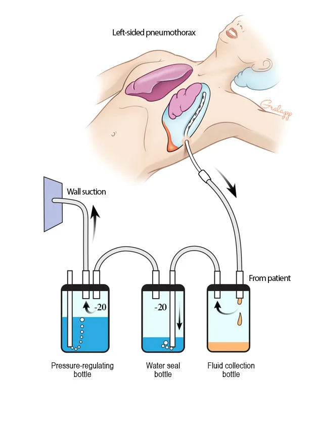
Securing the drain: To close the wound after the drain is withdrawn, a non-absorbable stitch and a loosely placed purse string should be used to hold the drain in place. An underwater seal is where the drain is fastened.
Care for chest drains: It is almost never advisable to clamp chest drains because this can cause a tension pneumthorax. The patient should never have the drain bottle raised above them, since this could cause fluid to spill into them. With every breath, the meniscus in the bottle ought to "swing."
Complications
harm to the upper abdominal, mediastinal, and thoracic tissues. The following conditions can occur: lung laceration, diaphragmatic injury, empyema and infection, blocked drain, recurrence after removal, and hemothorax (frequently an intercostal artery injury that may necessitate a thoracotomy).
Indication
Pneumothorax: Consistent or recurrent after simple aspiration; trauma; tension during needle decompression; ventilated patients.
Trauma in a hemothorax.
Pleural effusions, such as parapneumonic and malignant ones.
Postoperative: after, as in thoracotomy, oesophagectomy, and heart surgery.
Anatomy
The "safe triangle," which is formed by the anterior border of the latissimus dorsi, the lateral border of the pectoralis major, the apex below the axilla, and the upper border of the sixth rib, is where the chest drain insertion should be located. The diaphragm rises to the fifth rib upon expiration, hence placement should occur in the fourth or fifth rib gap. To prevent damage to the intercosal nerve and vascular bundles that run beneath each rib, the superior portion of the rib should be covered by the dissection and drain location.
Investigations
Tension pneumothorax: None is required; immediately do emergency decompression using a large-bore cannula in the midclavicular line's second intercostal gap.
Prior to procedure: Imaging: CXR, sometimes CT, depending on the situation being treated. In cases of effusions or empyemas, ultrasoundguided drain insertion can be helpful in guiding safe placement.
oxygen, analgesia, informed consent and explanation, and coagulopathy repair.
Post-procedure: To determine tube location, repeat CXR. close observation of production, bubbling, and swinging.
Actions
Positioning: To provide access to the sidewall of the chest, position the patient, for example, by reclining back with their arm abducted. Verify the right side. Clean the skin using aseptic approach, then drape.
Make an incision and inject a local anesthetic. Make an incision at the fourth or fifth intercostal space, usually just anterior to the midaxillary line. In order to prevent the neurovascular bundle from entering the chest through the pleura with a palpable "pop," bluntly dissect through the intercostal muscles above the fifth or sixth rib.
Insertion: If required, insert a chest drain (32–36F in trauma) fixed on a clamp and enter a finger to make sure there are no adhesions. Drainage is directed apically for pneumothorax and basally for fluid.
Securing the drain: To close the wound after the drain is withdrawn, a non-absorbable stitch and a loosely placed purse string should be used to hold the drain in place. An underwater seal is where the drain is fastened.
Care for chest drains: It is almost never advisable to clamp chest drains because this can cause a tension pneumthorax. The patient should never have the drain bottle raised above them, since this could cause fluid to spill into them. With every breath, the meniscus in the bottle ought to "swing."
Complications
harm to the upper abdominal, mediastinal, and thoracic tissues. The following conditions can occur: lung laceration, diaphragmatic injury, empyema and infection, blocked drain, recurrence after removal, and hemothorax (frequently an intercostal artery injury that may necessitate a thoracotomy).
0 Comments
