- Published on
Surgery - Cholangiocarcinoma
Introduction
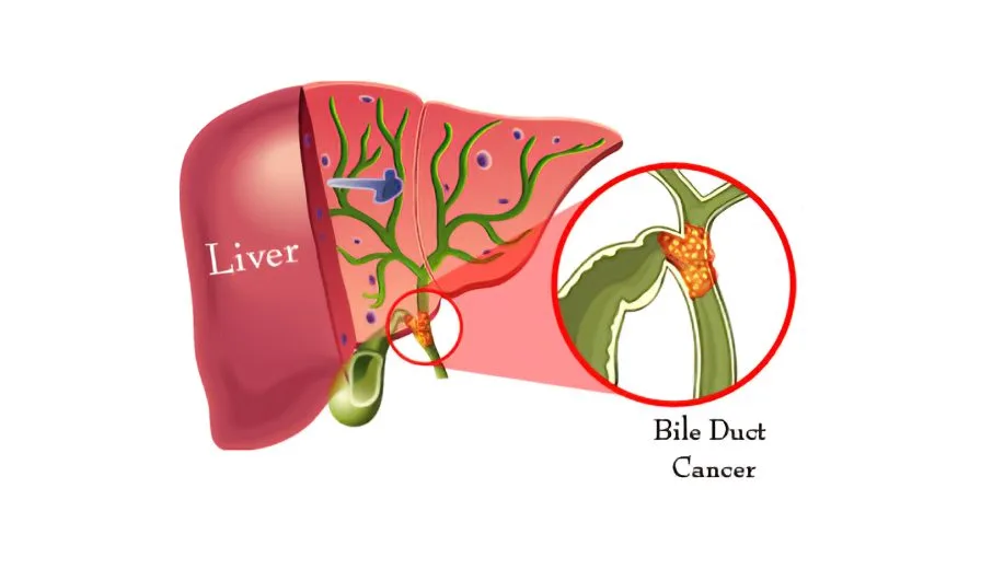
Epithelial cancer that starts in the bile ducts.
Etiology
Chronic inflammation and cholestasis are linked to various conditions such as primary sclerosing cholangitis (1.5% cumulative annual risk), choledochal cysts, congenital hepatic fibrosis, Thorotrast (a contrast agent used from the 1930s to 1950s), hepatolithiasis, parasitic infections of the biliary tract (e.g., Clonorchis sinensis, liver flukes), Lynch syndrome II, and biliary papillomatosis.
Epidemiology
Though uncommon (0.3–0.6% of cancer-related deaths), the incidence is rising and has surpassed hepatocellular carcinoma as the leading cause of primary liver tumor death in England. Asia has the highest incidence (caused by parasite infection). somewhat more prevalent in men.
History
Yellow skin and sclera, pale feces, black urine, and pruritus are symptoms of obstructive jaundice.
ache or fullness in the abdomen.
Malignancy symptoms include weariness, malaise, and weight loss.
Examining jaundice in detail. Palpable gallbladder (Courvoisier's law suggests that an enlarged gallbladder in the presence of jaundice is more likely to be caused by cancer of the pancreas or the lower biliary tree).
In more advanced cases, an upper quadrant mass or the epigastric area.
Pathogenesis
classified as extrahepatic [90%, hilar (Klatskin tumors), mid-duct, distal, and diffuse] or intrahepatic (10%) based on the location. Hilar tumors are categorized by Bismuth into categories I through V according to where they are in regard to the hepatic duct confluence. There are three distinct extrahepatic growth patterns: intraductal growing, periductal infiltrating, and bulk building.
Investigational studies
Blood: coagulation, FBC, U&Es, LFTs, and tumor markers (CEA and CA19-9, which are frequently elevated but not specific).
Endoscopy: ERCP/Endoscopic ultrasonography allows for the removal of obstructive jaundice, bile cytology, and tumor biopsies when available. An alternative for situations when bile duct access is challenging is percutaneous transhepatic cholangiography (PTC).
Ultrasound: Will demonstrate biliary channel dilatation, however sensitivity will vary.
To visualize any regional spread and stage the tumor, use CT, MRI, MRCP, or PET scanning.
When considering surgery, an arteriogram (invasive or MR) is crucial to demonstrate any involvement of the surrounding arterial systems.
TNM staging is used.
Management
Medical: Palliative treatments for tumors that cannot be removed. Metal or plastic stents are inserted during endoscopic (or percutaneous) biliary decompression; the latter have greater patency rates.
Chemo/Radiotherapy: Brachytherapy or intracavitary can shrink tumors, however currently, response rates to chemotherapy are not very good. It has been demonstrated that photodynamic therapy reduces symptoms and may increase survival.
Operative: The sole curative measure, yet less than 15% are treatable. Single intrahepatic tumors: lobectomy or segmentectomy. For hemar tumors, portal veinembolization is followed by an extensive hepatectomy. Whipple's surgery (proximal pancreaticoduodenectomy with choledocho- or hepaticojejunostomy) is used for distal tumors. Since non-curative/debulking surgery does not significantly improve survival, resection should only be done with the intention of curing. Liver transplant outcomes are not good.
Complications
cholangitis, obstructive jaundice, and metastases (local or lymphatic hepatic dissemination are common pathways).
Prognosis
Poor, less than a 24-month median survival. 40% of patients survive after five years after resection.
Introduction
Epithelial cancer that starts in the bile ducts.
Etiology
Chronic inflammation and cholestasis are linked to various conditions such as primary sclerosing cholangitis (1.5% cumulative annual risk), choledochal cysts, congenital hepatic fibrosis, Thorotrast (a contrast agent used from the 1930s to 1950s), hepatolithiasis, parasitic infections of the biliary tract (e.g., Clonorchis sinensis, liver flukes), Lynch syndrome II, and biliary papillomatosis.
Epidemiology
Though uncommon (0.3–0.6% of cancer-related deaths), the incidence is rising and has surpassed hepatocellular carcinoma as the leading cause of primary liver tumor death in England. Asia has the highest incidence (caused by parasite infection). somewhat more prevalent in men.
History
Yellow skin and sclera, pale feces, black urine, and pruritus are symptoms of obstructive jaundice.
ache or fullness in the abdomen.
Malignancy symptoms include weariness, malaise, and weight loss.
Examining jaundice in detail. Palpable gallbladder (Courvoisier's law suggests that an enlarged gallbladder in the presence of jaundice is more likely to be caused by cancer of the pancreas or the lower biliary tree).
In more advanced cases, an upper quadrant mass or the epigastric area.
Pathogenesis
classified as extrahepatic [90%, hilar (Klatskin tumors), mid-duct, distal, and diffuse] or intrahepatic (10%) based on the location. Hilar tumors are categorized by Bismuth into categories I through V according to where they are in regard to the hepatic duct confluence. There are three distinct extrahepatic growth patterns: intraductal growing, periductal infiltrating, and bulk building.
Investigational studies
Blood: coagulation, FBC, U&Es, LFTs, and tumor markers (CEA and CA19-9, which are frequently elevated but not specific).
Endoscopy: ERCP/Endoscopic ultrasonography allows for the removal of obstructive jaundice, bile cytology, and tumor biopsies when available. An alternative for situations when bile duct access is challenging is percutaneous transhepatic cholangiography (PTC).
Ultrasound: Will demonstrate biliary channel dilatation, however sensitivity will vary.
To visualize any regional spread and stage the tumor, use CT, MRI, MRCP, or PET scanning.
When considering surgery, an arteriogram (invasive or MR) is crucial to demonstrate any involvement of the surrounding arterial systems.
TNM staging is used.
Management
Medical: Palliative treatments for tumors that cannot be removed. Metal or plastic stents are inserted during endoscopic (or percutaneous) biliary decompression; the latter have greater patency rates.
Chemo/Radiotherapy: Brachytherapy or intracavitary can shrink tumors, however currently, response rates to chemotherapy are not very good. It has been demonstrated that photodynamic therapy reduces symptoms and may increase survival.
Operative: The sole curative measure, yet less than 15% are treatable. Single intrahepatic tumors: lobectomy or segmentectomy. For hemar tumors, portal veinembolization is followed by an extensive hepatectomy. Whipple's surgery (proximal pancreaticoduodenectomy with choledocho- or hepaticojejunostomy) is used for distal tumors. Since non-curative/debulking surgery does not significantly improve survival, resection should only be done with the intention of curing. Liver transplant outcomes are not good.
Complications
cholangitis, obstructive jaundice, and metastases (local or lymphatic hepatic dissemination are common pathways).
Prognosis
Poor, less than a 24-month median survival. 40% of patients survive after five years after resection.
0 Comments
