- Published on
Surgery - Colon Cancer
Introduction
Large bowel adenocarcinoma.
Etiology
Genetic and environmental variables have been linked. It is believed that a series of events, including the accumulation of genetic alterations, the activation of oncogenes (such APC and K-ras) and the inactivation of tumour suppressor genes (like p53 and DCC), leads from epithelial dysplasia to adenoma and ultimately carcinoma.
Risk Factors
polyps in the colon, a history of colorectal cancer, and inflammatory bowel disease, especially chronic ulcerative colitis. Lynch syndrome and familial adenomatous polyposis are two genetic syndromes linked to an increased risk.
Epidemiology
With 37,500 cases annually, cancer is the second cause of cancer death and the third most frequent cancer in the UK. 60–65 years old on average at diagnosis. Colon cancers: females > males, rectal carcinomas: males > females.
History
The location and size of the tumor affect the symptoms.
Through the NHS bowel cancer screening program, patients may come with positive faecal occult blood tests even when they are asymptomatic.
colon and rectum on the left side: altered bowel habits, rectal hemorrhage, or blood or mucus mixed with feces. Tenesmus, or the sense of incomplete emptying following bowel movement, is another symptom of rectal masses.
Right-sided colon: Later presentation characterized by anemia symptoms, weight loss, general malaise, or, in rare cases, lower abdominal pain.
About 20% of tumors will cause pain and distension in an emergency room due to major intestinal blockage, bleeding, or peritonitis from a perforation.
Examination
There might not be any indicators.
Particularly in cases of right-sided lesions, abdominal mass, hepatomegaly, metastatic illness, and "shifting dullness" of ascites, anemia may be the only symptom.
During a rectal examination, low-lying rectal tumors may feel palpable.
Pathogenesis
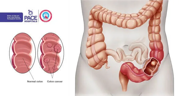
30% of the colon is in the ascending colon, 60% is in the rectum and sigmoid colon, and the remaining 40% is in the descending and transverse colon. Tumors can produce polypoid, exophytic masses or be annular, resembling "apple core" lesions. Duke and the TNM system are examples of staging systems.
Investigations
Blood: tumor markers (CEA to track therapy response or disease recurrence), LFT, and FBC (for anemia).
Blood in the stool: This test can be used to screen for occult or frank blood.
Endoscopy: colonoscopy and sigmoidoscopy. allows for biopsy and visualization. Isolated small carcinoma in situ lesions may be treated with polypectomy or endoscopic mucosal excision.
Imaging methods include endorectal ultrasonography, PET scanning, CT, MRI, and barium enema.
Management
By identifying and treating pre-malignant polyps and early illness, screening lowers mortality.
Operation: Depending on the stage and location, for example. Caecal tumors: Hemicilocectomy to the right. Right hemicolectomy extended for transverse colon tumors.
Left hemicolectomy for colon tumors that descend.
Sigmoid tumors: Resection anteriorly.
High mid-rectal: complete mesorectal excision combined with anterior resection.
If the tumor cannot be cleared, low rectal: abdominoperineal resection.
Emergency: Depending on the location and clinical presentation; examples include removal of a tumor, Hartmann's surgery, primary anastomosis, and a malfunctioning ileostomy.
Radiotherapy can be administered as adjuvant therapy to lower the chance of a local recurrence or as a neoadjuvant setting to downstage rectal tumors before resection.
Chemotherapy: For metastatic diseases or as an adjuvant treatment. 5-fluorouracil combination chemotherapy regimens are prevalent (e.g. FOLFOX). In cases of metastatic disease, chemotherapy is combined with bevacizumab (anti-vascular endothelial growth factor) or cetuximab (anti-epidermal growth factor receptor in the absence of a K-ras mutation).
Complications
Metastatic illness, recurrence, fistula formation, obstruction or perforation of the bowel.
Prognosis
varies according on the stage. It has been demonstrated that in situations that are treatable, those with poor mismatch repair have better outcomes.
Dukes Spread and 5-year survival extent:
An 90%–95% confined to the intestinal wall 77% C of serosa breached but no lymph nodes were affected serosa breached, including 48% of the lymph nodes D distant liver metastases The original Duke's staging does not include the 5–10% D stage.
Introduction
Large bowel adenocarcinoma.
Etiology
Genetic and environmental variables have been linked. It is believed that a series of events, including the accumulation of genetic alterations, the activation of oncogenes (such APC and K-ras) and the inactivation of tumour suppressor genes (like p53 and DCC), leads from epithelial dysplasia to adenoma and ultimately carcinoma.
Risk Factors
polyps in the colon, a history of colorectal cancer, and inflammatory bowel disease, especially chronic ulcerative colitis. Lynch syndrome and familial adenomatous polyposis are two genetic syndromes linked to an increased risk.
Epidemiology
With 37,500 cases annually, cancer is the second cause of cancer death and the third most frequent cancer in the UK. 60–65 years old on average at diagnosis. Colon cancers: females > males, rectal carcinomas: males > females.
History
The location and size of the tumor affect the symptoms.
Through the NHS bowel cancer screening program, patients may come with positive faecal occult blood tests even when they are asymptomatic.
colon and rectum on the left side: altered bowel habits, rectal hemorrhage, or blood or mucus mixed with feces. Tenesmus, or the sense of incomplete emptying following bowel movement, is another symptom of rectal masses.
Right-sided colon: Later presentation characterized by anemia symptoms, weight loss, general malaise, or, in rare cases, lower abdominal pain.
About 20% of tumors will cause pain and distension in an emergency room due to major intestinal blockage, bleeding, or peritonitis from a perforation.
Examination
There might not be any indicators.
Particularly in cases of right-sided lesions, abdominal mass, hepatomegaly, metastatic illness, and "shifting dullness" of ascites, anemia may be the only symptom.
During a rectal examination, low-lying rectal tumors may feel palpable.
Pathogenesis
30% of the colon is in the ascending colon, 60% is in the rectum and sigmoid colon, and the remaining 40% is in the descending and transverse colon. Tumors can produce polypoid, exophytic masses or be annular, resembling "apple core" lesions. Duke and the TNM system are examples of staging systems.
Investigations
Blood: tumor markers (CEA to track therapy response or disease recurrence), LFT, and FBC (for anemia).
Blood in the stool: This test can be used to screen for occult or frank blood.
Endoscopy: colonoscopy and sigmoidoscopy. allows for biopsy and visualization. Isolated small carcinoma in situ lesions may be treated with polypectomy or endoscopic mucosal excision.
Imaging methods include endorectal ultrasonography, PET scanning, CT, MRI, and barium enema.
Management
By identifying and treating pre-malignant polyps and early illness, screening lowers mortality.
Operation: Depending on the stage and location, for example. Caecal tumors: Hemicilocectomy to the right. Right hemicolectomy extended for transverse colon tumors.
Left hemicolectomy for colon tumors that descend.
Sigmoid tumors: Resection anteriorly.
High mid-rectal: complete mesorectal excision combined with anterior resection.
If the tumor cannot be cleared, low rectal: abdominoperineal resection.
Emergency: Depending on the location and clinical presentation; examples include removal of a tumor, Hartmann's surgery, primary anastomosis, and a malfunctioning ileostomy.
Radiotherapy can be administered as adjuvant therapy to lower the chance of a local recurrence or as a neoadjuvant setting to downstage rectal tumors before resection.
Chemotherapy: For metastatic diseases or as an adjuvant treatment. 5-fluorouracil combination chemotherapy regimens are prevalent (e.g. FOLFOX). In cases of metastatic disease, chemotherapy is combined with bevacizumab (anti-vascular endothelial growth factor) or cetuximab (anti-epidermal growth factor receptor in the absence of a K-ras mutation).
Complications
Metastatic illness, recurrence, fistula formation, obstruction or perforation of the bowel.
Prognosis
varies according on the stage. It has been demonstrated that in situations that are treatable, those with poor mismatch repair have better outcomes.
Dukes Spread and 5-year survival extent:
An 90%–95% confined to the intestinal wall 77% C of serosa breached but no lymph nodes were affected serosa breached, including 48% of the lymph nodes D distant liver metastases The original Duke's staging does not include the 5–10% D stage.
0 Comments
