- Published on
Surgery - Cord Compression and Injury
Introduction
damage to the spinal cord, the location and severity of which will determine the neurological symptoms. Acute compression of the cord is urgent.
Etiology
Direct trauma-related contusion to the spinal cord;-compression due to bone/disc fragmentation or trauma-related hemorrhage;-compression due to extrinsic lesions, such as: disc protrusion; tumors (primary, secondary, or myeloma); spinal abscesses (such as TB and staphylococcus) abnormalities of the spinal vessels
Epidemiology
Typical. All age groups experience trauma. In senior age groups, cancer and disc disease are more prevalent.
History
Past medical history of trauma, illness, or damage.
weakness, pain, and loss of senses.
interruption of bladder or bowel function. impotence.
Urinary retention, saddle anesthesia, and bilateral sciatica are symptoms of cauda-equina syndrome.
Seen in spinal cord hemisection: Brown-Sequard syndrome. ipsilateral leg weakness and contralateral leg numbness.
Examination
Take a close look at both the motor and sensory levels.
Trauma to the spinal cord: Early hyperacute symptoms include diaphragmatic breathing, diminished anal tone, hyporeflexia, priapism, and spinal shock (# BP without tachycardia).
Flaccid paraparesis, urine retention, decreased anal tone, saddle anesthesia, and compromised knee, ankle, and bulbocavernous reflexes are all symptoms of cauda-equina syndrome. Perhaps not symmetrical.
Urinary retention combined with flaccid and spastic paraparesis is known as Conus medullaris syndrome.
Below the lesion, hypertonicity and hyperreflexia are present. Sensory disruption is often symmetrical and distributed perianally.
Sequard-Brown syndrome: observed in spinal cord hemisection. Ipsilateral spastic paralysis, loss of postural sensation, and contralateral loss of pain and temperature sense occur below the level of the lesion.
Differentiate between radiculopathies and cord compression: Radiculopathies are brought on by compression of the nerve root at the exit foramina or in the spinal canal. Only at that motor and sensory level does this result in LMN damage; beyond that level, no UMN indications are present.
Motor
C3–C5: Diaphragm L2: Hip flexion
C5: Shoulder abduction. L3: Knee extension
C6: Forearm flexion L4: Ankle dorsiflexion
C7: Forearm extension L5: Big toe extension
C8: Wrist/finger flexion S1: Ankle plantar flexion
T1: Finger abduction
Reflexes
C5–C6: Biceps reflex L1–L2: Cremaster reflex
C6: Brachioradialis (supinator) reflex L3–L4: Knee reflex
C7: Triceps reflex S1–S2: Ankle reflex
Hoffmann/s sign: UMN in upper limb S2–S4: Anocutaneous refl
S2–S4: Bulbocavernous reflex
Sensory
C4: Supraclavicular fossa T10: Umbilicus
C5: Clavicles T12: Hip girdle
C6: Thumb L4: Medial malleolus
C7: Middle finger S1: Lateral malleolus
C8: Little finger S2: Scrotum
T4: Nipples S3–S5: Perianal region
Investigations
Trauma radiology includes AP and lateral spine (also known as peg view), thoracic, and lumbar radiographs. High-resolution CT scans of the spine, which enable formatting for 3D visualization, are gradually replacing them.
Spine emergency MRI: To see the cord and soft tissue. Changes in cord signal and canal stenosis are important indicators.
Blood: protein electrophoresis, Ca2+, ESR, bone profile, FBC, and U&E.
Bence Jones protein in the urine is a sign of multiple myeloma.
Management
Traumatic cord injury: The ATLS recommendations should be followed while managing trauma injuries.
While it is debatable, NASCIS trial data suggests that giving high-dose steroids (methylprednisolone) to patients with severe spinal cord injuries for less than eight hours can improve their motor function.
Cord compression: If a tumor is found after imaging, high-dose steroids, such as dexamethasone, should be administered as away to lessen cord compression. Emergency radiation may potentially be effective in treating tumors. Get guidance from a neurosurgeon.
Surgery: Often required to remove the origin of compression and/or relieve compression.
External surgical stabilization: Using a halo or traction device, for instance.
Internal surgical stabilization: Using metal implants or bone grafts, for instance.
Surgical decompression: To relieve compression, a laminectomy, for instance.
For disc prolapses, a discectomy or microdiscectomy is used.
Rehabilitation: A multidisciplinary rehabilitation program including occupational therapy and physiotherapy, ideally in a specialized spinal unit.
Complications
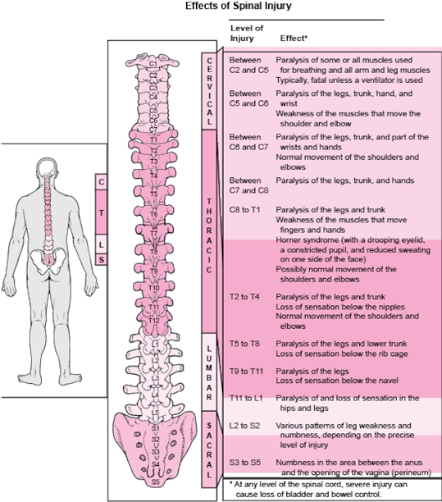
Spinal function below the lesion is lost in cases of severe injury.
Paralysis of the breathing system: lesion above C4.
Quadriplegia, C4–T1.
Mid-thoracic: If above T6, paraplegia and autonomic dysreflexia.
S1: Loss of rectum and bladder control due to sacral parasympathetic loss.
Immobility-related complications include pressure sores, DVT, long-term spasticity (posing a risk of deformity), and heterotopic ossification.
Prognosis
When converted to the Frankel grade, the American Spinal Injury Association (ASIA) Impairment Scale after 72 hours after the injury has prognostic significance (see www.asia-spinalinjury.org for score sheet). Depending on the extent of the spinal cord injury, there may be some improvement over time.
Introduction
damage to the spinal cord, the location and severity of which will determine the neurological symptoms. Acute compression of the cord is urgent.
Etiology
Direct trauma-related contusion to the spinal cord;-compression due to bone/disc fragmentation or trauma-related hemorrhage;-compression due to extrinsic lesions, such as: disc protrusion; tumors (primary, secondary, or myeloma); spinal abscesses (such as TB and staphylococcus) abnormalities of the spinal vessels
Epidemiology
Typical. All age groups experience trauma. In senior age groups, cancer and disc disease are more prevalent.
History
Past medical history of trauma, illness, or damage.
weakness, pain, and loss of senses.
interruption of bladder or bowel function. impotence.
Urinary retention, saddle anesthesia, and bilateral sciatica are symptoms of cauda-equina syndrome.
Seen in spinal cord hemisection: Brown-Sequard syndrome. ipsilateral leg weakness and contralateral leg numbness.
Examination
Take a close look at both the motor and sensory levels.
Trauma to the spinal cord: Early hyperacute symptoms include diaphragmatic breathing, diminished anal tone, hyporeflexia, priapism, and spinal shock (# BP without tachycardia).
Flaccid paraparesis, urine retention, decreased anal tone, saddle anesthesia, and compromised knee, ankle, and bulbocavernous reflexes are all symptoms of cauda-equina syndrome. Perhaps not symmetrical.
Urinary retention combined with flaccid and spastic paraparesis is known as Conus medullaris syndrome.
Below the lesion, hypertonicity and hyperreflexia are present. Sensory disruption is often symmetrical and distributed perianally.
Sequard-Brown syndrome: observed in spinal cord hemisection. Ipsilateral spastic paralysis, loss of postural sensation, and contralateral loss of pain and temperature sense occur below the level of the lesion.
Differentiate between radiculopathies and cord compression: Radiculopathies are brought on by compression of the nerve root at the exit foramina or in the spinal canal. Only at that motor and sensory level does this result in LMN damage; beyond that level, no UMN indications are present.
Motor
C3–C5: Diaphragm L2: Hip flexion
C5: Shoulder abduction. L3: Knee extension
C6: Forearm flexion L4: Ankle dorsiflexion
C7: Forearm extension L5: Big toe extension
C8: Wrist/finger flexion S1: Ankle plantar flexion
T1: Finger abduction
Reflexes
C5–C6: Biceps reflex L1–L2: Cremaster reflex
C6: Brachioradialis (supinator) reflex L3–L4: Knee reflex
C7: Triceps reflex S1–S2: Ankle reflex
Hoffmann/s sign: UMN in upper limb S2–S4: Anocutaneous refl
S2–S4: Bulbocavernous reflex
Sensory
C4: Supraclavicular fossa T10: Umbilicus
C5: Clavicles T12: Hip girdle
C6: Thumb L4: Medial malleolus
C7: Middle finger S1: Lateral malleolus
C8: Little finger S2: Scrotum
T4: Nipples S3–S5: Perianal region
Investigations
Trauma radiology includes AP and lateral spine (also known as peg view), thoracic, and lumbar radiographs. High-resolution CT scans of the spine, which enable formatting for 3D visualization, are gradually replacing them.
Spine emergency MRI: To see the cord and soft tissue. Changes in cord signal and canal stenosis are important indicators.
Blood: protein electrophoresis, Ca2+, ESR, bone profile, FBC, and U&E.
Bence Jones protein in the urine is a sign of multiple myeloma.
Management
Traumatic cord injury: The ATLS recommendations should be followed while managing trauma injuries.
While it is debatable, NASCIS trial data suggests that giving high-dose steroids (methylprednisolone) to patients with severe spinal cord injuries for less than eight hours can improve their motor function.
Cord compression: If a tumor is found after imaging, high-dose steroids, such as dexamethasone, should be administered as away to lessen cord compression. Emergency radiation may potentially be effective in treating tumors. Get guidance from a neurosurgeon.
Surgery: Often required to remove the origin of compression and/or relieve compression.
External surgical stabilization: Using a halo or traction device, for instance.
Internal surgical stabilization: Using metal implants or bone grafts, for instance.
Surgical decompression: To relieve compression, a laminectomy, for instance.
For disc prolapses, a discectomy or microdiscectomy is used.
Rehabilitation: A multidisciplinary rehabilitation program including occupational therapy and physiotherapy, ideally in a specialized spinal unit.
Complications
Spinal function below the lesion is lost in cases of severe injury.
Paralysis of the breathing system: lesion above C4.
Quadriplegia, C4–T1.
Mid-thoracic: If above T6, paraplegia and autonomic dysreflexia.
S1: Loss of rectum and bladder control due to sacral parasympathetic loss.
Immobility-related complications include pressure sores, DVT, long-term spasticity (posing a risk of deformity), and heterotopic ossification.
Prognosis
When converted to the Frankel grade, the American Spinal Injury Association (ASIA) Impairment Scale after 72 hours after the injury has prognostic significance (see www.asia-spinalinjury.org for score sheet). Depending on the extent of the spinal cord injury, there may be some improvement over time.
0 Comments
