- Published on
Surgery - Deep Vein Thrombosis
Introduction
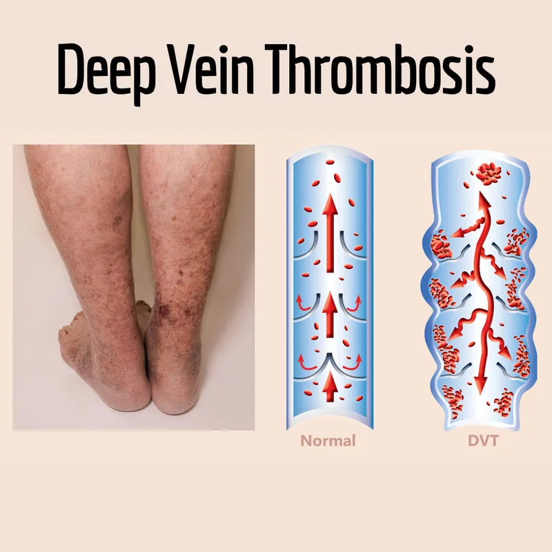
thrombus formation in the deep veins, usually in the thigh or calf.
Etiology
Virchow's triad: blood hypercoagulability, vascular wall damage, and venous stasis.
Risk Factors
Factor V Leiden, deficits in Protein C or Protein S deficienceis Prothrombin mutation, and insufficient Antithrombin III are all inherited.
Oral contraceptive pill, heparin-induced thrombocytopenia, immobility, obesity, pregnancy, polycythemia, anti-phospholipid syndrome, smoking, nephrotic syndrome, and aggressive cancer are among the acquired conditions.
Epidemiology
Frequent, particularly in hospitalized patients; precise prevalence is unclear. Venous insufficiency and ulceration, long-term consequences of DVT, afflict 0.5 percent of the population. Around 145 per 100,000, around.
History
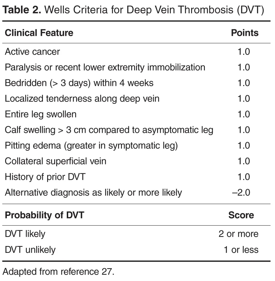
asymptomatic or soreness or edema in the lower limbs. may exhibit pulmonary embolus signs and symptoms.
Examination
Check for any swelling or soreness in the calf.
It is uncommon to have severe leg cyanosis and oedema (phlegmasia cerulea dolens).
checking the airways for indications of a pulmonary embolus.
Investigations
Ultrasound Doppler: The gold standard. Less sensitive in calf veins, but good sensitivity in femoral veins.
Bloods: Only low-risk individuals can benefit from the sensitive but very non-specific D-dimers (fibrinogen degradation products) as a negative predictor. Before beginning anticoagulation, a thrombophilia screen should be sent if necessary (e.g., recurring occurrences).
FBC (platelet count before beginning heparin), coagulation, and U&E.
ABG, CXR, and ECG: If there is a possibility that PE may be present.
Management
Anticoagulation: While waiting for a therapeutic INR from warfarin anticoagulation, patients should receive heparin treatment. Anticoagulation is prescribed for three months for DVTs that do not extend above the knee and for six months for those that do. Warfarin may be needed for a long time if DVTs recur. Interventional radiology should install an IVC filter, such as the Greenfield filter, if active anticoagulation is contraindicated or there is a high risk of embolization in order to prevent embolus to the lungs.
Using graded compression stockings is one way to prevent. mobilizing, if at all possible. Hospital patients who are considered to be at-risk should receive preventive heparin, such as low-molecular-weight heparin, provided there are no contraindications.
Complications
Disease symptoms include pulmonary embolus, impaired vein valves, and persistent lower limb venous insufficiency (post-thrombotic syndrome). Venous infarction, or phlegmasia cereulea dolens, is uncommon.
Heparin-induced thrombocytopenia and bleeding are side effects of treatment.
Prognosis
based on the severity of DVT. More proximal DVTs have a higher chance of propagation and embolization, which, if significant, may be deadly. Below-knee DVTs have a decreased risk of embolus.
Introduction
thrombus formation in the deep veins, usually in the thigh or calf.
Etiology
Virchow's triad: blood hypercoagulability, vascular wall damage, and venous stasis.
Risk Factors
Factor V Leiden, deficits in Protein C or Protein S deficienceis Prothrombin mutation, and insufficient Antithrombin III are all inherited.
Oral contraceptive pill, heparin-induced thrombocytopenia, immobility, obesity, pregnancy, polycythemia, anti-phospholipid syndrome, smoking, nephrotic syndrome, and aggressive cancer are among the acquired conditions.
Epidemiology
Frequent, particularly in hospitalized patients; precise prevalence is unclear. Venous insufficiency and ulceration, long-term consequences of DVT, afflict 0.5 percent of the population. Around 145 per 100,000, around.
History
asymptomatic or soreness or edema in the lower limbs. may exhibit pulmonary embolus signs and symptoms.
Examination
Check for any swelling or soreness in the calf.
It is uncommon to have severe leg cyanosis and oedema (phlegmasia cerulea dolens).
checking the airways for indications of a pulmonary embolus.
Investigations
Ultrasound Doppler: The gold standard. Less sensitive in calf veins, but good sensitivity in femoral veins.
Bloods: Only low-risk individuals can benefit from the sensitive but very non-specific D-dimers (fibrinogen degradation products) as a negative predictor. Before beginning anticoagulation, a thrombophilia screen should be sent if necessary (e.g., recurring occurrences).
FBC (platelet count before beginning heparin), coagulation, and U&E.
ABG, CXR, and ECG: If there is a possibility that PE may be present.
Management
Anticoagulation: While waiting for a therapeutic INR from warfarin anticoagulation, patients should receive heparin treatment. Anticoagulation is prescribed for three months for DVTs that do not extend above the knee and for six months for those that do. Warfarin may be needed for a long time if DVTs recur. Interventional radiology should install an IVC filter, such as the Greenfield filter, if active anticoagulation is contraindicated or there is a high risk of embolization in order to prevent embolus to the lungs.
Using graded compression stockings is one way to prevent. mobilizing, if at all possible. Hospital patients who are considered to be at-risk should receive preventive heparin, such as low-molecular-weight heparin, provided there are no contraindications.
Complications
Disease symptoms include pulmonary embolus, impaired vein valves, and persistent lower limb venous insufficiency (post-thrombotic syndrome). Venous infarction, or phlegmasia cereulea dolens, is uncommon.
Heparin-induced thrombocytopenia and bleeding are side effects of treatment.
Prognosis
based on the severity of DVT. More proximal DVTs have a higher chance of propagation and embolization, which, if significant, may be deadly. Below-knee DVTs have a decreased risk of embolus.
0 Comments
