- Published on
Surgery - Extradural Hemorrhage
Introduction
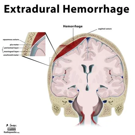
blood clotting and seeping into the extradural area.
Etiology
Because the squamous temporal bone is the thinnest portion of the cranial vault, head trauma often results in a fracture that can rupture the middle meningeal artery. Rapid blood clotting from arterial bleeding removes the dura from the skull's inner table. As a result, the underlying brain parenchyma is compressed and the ICP is elevated.
Epidemiology
In the UK, there is an annual incidence of 20/100,000 severe head injuries, or 10%. Mostly observed in young adults. Not frequently seen in older adults (subdural hemorrhages are more common in this age group).
History
Head injury with a momentary loss of consciousness, followed by a lucid interval, then development of steady degradation in conscious level.
Examination
symptoms of a broken or injured scalp.
ache.
declining GCS.
indications of an elevated ICP, such as a dilated, unresponsive pupil on the side of the wound.
Cushing's sign (increasing blood pressure and bradycardia) and abnormal posturing (decorticate and decerebrate) are examples of late indications.
Investigations
Quick CT scan: Identifies the site of the hemorrhage and is diagnostic. A haematoma that is convex or lens-shaped results from an arterial bleed. Midline shift, ventricular compression, basal cistern obliteration, and sulcal effacement are indications of elevated ICP.
Management
Early head injury management: To establish ABC and cervical spine control, according to ATLS standards. After the patient is stabilized, a CT scan is quickly performed to determine the extent of the brain injuries.
Surgical: Decompressive evacuation of the hemogloba by diathermy or cutting off the bleeding source in an emergency craniotomy. For postop monitoring, an ICP monitor could be installed. Supportive care and close observation are necessary, frequently in an ICU situation.
Complications
Acutely, brain herniation and mortality provide the most risk.
Amnesia, cognitive disability, and post-traumatic brain injury may be related in the long run.
Prognosis
Mortality rates are correlated with intracerebral injuries and the initial GCS. The prognosis is good if treatment is received early, and the underlying brain injury is typically mild.
Introduction
blood clotting and seeping into the extradural area.
Etiology
Because the squamous temporal bone is the thinnest portion of the cranial vault, head trauma often results in a fracture that can rupture the middle meningeal artery. Rapid blood clotting from arterial bleeding removes the dura from the skull's inner table. As a result, the underlying brain parenchyma is compressed and the ICP is elevated.
Epidemiology
In the UK, there is an annual incidence of 20/100,000 severe head injuries, or 10%. Mostly observed in young adults. Not frequently seen in older adults (subdural hemorrhages are more common in this age group).
History
Head injury with a momentary loss of consciousness, followed by a lucid interval, then development of steady degradation in conscious level.
Examination
symptoms of a broken or injured scalp.
ache.
declining GCS.
indications of an elevated ICP, such as a dilated, unresponsive pupil on the side of the wound.
Cushing's sign (increasing blood pressure and bradycardia) and abnormal posturing (decorticate and decerebrate) are examples of late indications.
Investigations
Quick CT scan: Identifies the site of the hemorrhage and is diagnostic. A haematoma that is convex or lens-shaped results from an arterial bleed. Midline shift, ventricular compression, basal cistern obliteration, and sulcal effacement are indications of elevated ICP.
Management
Early head injury management: To establish ABC and cervical spine control, according to ATLS standards. After the patient is stabilized, a CT scan is quickly performed to determine the extent of the brain injuries.
Surgical: Decompressive evacuation of the hemogloba by diathermy or cutting off the bleeding source in an emergency craniotomy. For postop monitoring, an ICP monitor could be installed. Supportive care and close observation are necessary, frequently in an ICU situation.
Complications
Acutely, brain herniation and mortality provide the most risk.
Amnesia, cognitive disability, and post-traumatic brain injury may be related in the long run.
Prognosis
Mortality rates are correlated with intracerebral injuries and the initial GCS. The prognosis is good if treatment is received early, and the underlying brain injury is typically mild.
0 Comments
