- Published on
Surgery - Gastrointestinal Perforation
Introduction
GI tract wall perforation resulting in bowel contents spilling out.
Etiology
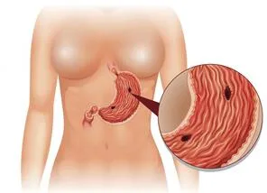
Perforated duodenum or gastric ulcers are the most frequent gastroduodenal conditions; gastric cancer (1-2%) is less common.
In the large intestine, diverticulitis and colon cancer account for the majority of cases (80%); an appendix perforation is a common consequence of appendicitis. Additional conditions include trauma, radiation enteritis, volvulus, ulcerative colitis (toxic megacolon), post-operative anastomotic leaks, and colonoscopy complications.
Rarely, small bowel: radiation enteritis, lymphoma, Crohn's disease, infection (typhoid, TB), trauma.
Boerhaave's syndrome (oesophagus): refer to oesophageal perforation. Iatrogenic perforation happens more frequently during stricture dilatation than it does during OGD.
Epidemiology
Cause determines incidence. On the other hand, bowel perforation-related stomach pain is a somewhat common and potentially fatal emergency.
History
Depending on the reason. Abdominal pain is generally linked to nausea and vomiting, with a fast onset.
Examination
The patient is ill, exhibiting symptoms of either localized or generalized peritonitis, including diminished or absent bowel sounds, guarding, and rigidity in the abdomen. Overlying gas causes the loss of liver dullness. pallor, pyrexia, shock, and dehydration symptoms.
Investigational studies
Blood: ABGs, coagulation, amylase (levels may be elevated in perforation), FBC, U&Es, and LFT.
Erect CXR: In 70% of patients, a perforated peptic ulcer will show gas behind the diaphragm (see Fig. 4).
AXR: Tissues may exhibit aberrant gas shadows. Gas on each side of the colon wall is referred to as Rigler's sign; alternatively, intraperitoneal gas can be seen on a lateral decubitus film.
CT scan: May identify underlying pathology and is extremely sensitive for free intraperitoneal gas.
Management
Intravenous rehydration, correction of electrolyte imbalances, broad-spectrum IV antibiotics, analgesics, urine catheter, and central line as needed are all part of resuscitation.
Conservative: Saved for patients with few symptoms, little contamination, or a high risk of anesthesia. In order to treat gastroduodenal perforations, high-dose PPIs, IV fluids, antibiotics, NG tubes, and monitoring are all recommended.
Operative: Gastroduodenal: Peritoneal lavage and laparoscopy or laparotomy An omental patch is applied once the puncture is sealed. A biopsy of a stomach ulcer is necessary to check for malignancy. Although gastroduodenal anastomosis and Billroth I partial gastrectomy can be performed, closure is more challenging than duodenal ulcers. If positive, post-operative elimination of Helicobacter pylori.
Large bowel: Perineural lavage and site of perforation detection via laparoscopy or laparotomy. removal of the affected colon, typically as a part of a Hartmann's procedure, along with the creation of an end colostomy, exteriorization as a mucous fistula, and closure of the distal stump. Resection and primary anastomosis combined with a failing ileostomy are the alternative. Resection and a primary anastomosis may be possible with a right colon perforation. A subtotal colectomy with a terminal ileostomy and preservation of the rectal stump is done in cases with toxic megacolon of ulcerative colitis (allows future repair of ileoanal pouch).
Complications
mortality, fistula development, peritonitis, and sepsis.
Prognosis
Gastroduodenal: Perforated gastric ulcers have a higher morbidity and death rate than duodenal ulcers, and the prognosis for perforated gastric carcinomas is extremely bad.
Greater prognosis with localized or restricted contamination in the large bowel. The mortality rate from fecal peritonitis is more than 50%.
Introduction
GI tract wall perforation resulting in bowel contents spilling out.
Etiology
Perforated duodenum or gastric ulcers are the most frequent gastroduodenal conditions; gastric cancer (1-2%) is less common.
In the large intestine, diverticulitis and colon cancer account for the majority of cases (80%); an appendix perforation is a common consequence of appendicitis. Additional conditions include trauma, radiation enteritis, volvulus, ulcerative colitis (toxic megacolon), post-operative anastomotic leaks, and colonoscopy complications.
Rarely, small bowel: radiation enteritis, lymphoma, Crohn's disease, infection (typhoid, TB), trauma.
Boerhaave's syndrome (oesophagus): refer to oesophageal perforation. Iatrogenic perforation happens more frequently during stricture dilatation than it does during OGD.
Epidemiology
Cause determines incidence. On the other hand, bowel perforation-related stomach pain is a somewhat common and potentially fatal emergency.
History
Depending on the reason. Abdominal pain is generally linked to nausea and vomiting, with a fast onset.
Examination
The patient is ill, exhibiting symptoms of either localized or generalized peritonitis, including diminished or absent bowel sounds, guarding, and rigidity in the abdomen. Overlying gas causes the loss of liver dullness. pallor, pyrexia, shock, and dehydration symptoms.
Investigational studies
Blood: ABGs, coagulation, amylase (levels may be elevated in perforation), FBC, U&Es, and LFT.
Erect CXR: In 70% of patients, a perforated peptic ulcer will show gas behind the diaphragm (see Fig. 4).
AXR: Tissues may exhibit aberrant gas shadows. Gas on each side of the colon wall is referred to as Rigler's sign; alternatively, intraperitoneal gas can be seen on a lateral decubitus film.
CT scan: May identify underlying pathology and is extremely sensitive for free intraperitoneal gas.
Management
Intravenous rehydration, correction of electrolyte imbalances, broad-spectrum IV antibiotics, analgesics, urine catheter, and central line as needed are all part of resuscitation.
Conservative: Saved for patients with few symptoms, little contamination, or a high risk of anesthesia. In order to treat gastroduodenal perforations, high-dose PPIs, IV fluids, antibiotics, NG tubes, and monitoring are all recommended.
Operative: Gastroduodenal: Peritoneal lavage and laparoscopy or laparotomy An omental patch is applied once the puncture is sealed. A biopsy of a stomach ulcer is necessary to check for malignancy. Although gastroduodenal anastomosis and Billroth I partial gastrectomy can be performed, closure is more challenging than duodenal ulcers. If positive, post-operative elimination of Helicobacter pylori.
Large bowel: Perineural lavage and site of perforation detection via laparoscopy or laparotomy. removal of the affected colon, typically as a part of a Hartmann's procedure, along with the creation of an end colostomy, exteriorization as a mucous fistula, and closure of the distal stump. Resection and primary anastomosis combined with a failing ileostomy are the alternative. Resection and a primary anastomosis may be possible with a right colon perforation. A subtotal colectomy with a terminal ileostomy and preservation of the rectal stump is done in cases with toxic megacolon of ulcerative colitis (allows future repair of ileoanal pouch).
Complications
mortality, fistula development, peritonitis, and sepsis.
Prognosis
Gastroduodenal: Perforated gastric ulcers have a higher morbidity and death rate than duodenal ulcers, and the prognosis for perforated gastric carcinomas is extremely bad.
Greater prognosis with localized or restricted contamination in the large bowel. The mortality rate from fecal peritonitis is more than 50%.
0 Comments
