- Published on
Surgery - Gastrointestinal Stromal Tumors
Introduction
Mesenchymal tumors of the intestines that could be produced from Cajal interstitial cells or pacemaker cells connected to the Auerbach's plexus that regulate peristalsis. Display an array of malignant potential ranging from extremely low risk to overtly malignant.
Etiology
typified by mutations in either PDGFR-a (platelet-derived growth factor receptor alpha, 5–10%) or KIT (75–80%, CD117), which together activate receptor tyrosine kinase signaling pathways and cause cell division.
Risk Factors
Neurofibromatosis type I, familial GIST syndrome, and Carney's triad.
Epidemiology
Annually, there are 11–15 cases per million and 129 cases of prevalence. Male to female, a large age range, with 75% of respondents being over 50 (median age 58).
History
Possible asymptomatic or inadvertent detection during imaging, laparotomy, or endoscopy. GI bleeding (70%) is one of the other manifestations, along with bloating and abdominal pain (57%), bowel blockage (30%), satiety and weight loss (22%), palpable mass (13%), and, less frequently, rupture.
Examination
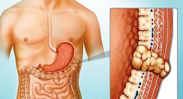
Results vary depending on the site, complexity, and size. can occur anywhere in the gastrointestinal system, with the stomach accounting for 50% of cases, small bowel for 25%, colon/rectum for 10%, mesentery, omentum and retroperitoneum for 10%, and oesophagus for 5%.
Investigational studies
Endoscopy: May show a submucosal tumor; biopsies are often negative unless they are "inkwell" biopsies. Endoscopic ultrasound: a traditionally hypoechoic mass that is next to the propria or muscularis mucosa. In general, perioperative or percutaneous biopsy is not advised due to the possibility of tumor rupture and dissemination.
Imaging: 18FDG PET scanning and CT abdominal imaging. in order to pinpoint the tumor's location.
Immunohistochemistry: CD34 (60–70%) and KIT (95%), positive.
Management
The preferred course of treatment for non-metastatic GISTs is surgical resection. If at all possible, complete excision should be tried as it provides a strong probability of recovery. Lymphadenectomy on a routine basis is not advised. For small-to-intermediate tumors, laparoscopic excision is an option. Adjuvant imatinib and neoadjuvent are undergoing clinical studies.
advanced illness To treat advanced or metastatic GISTs, imatinib binds competitively to the ATP binding site and inhibits the receptor tyrosine kinases KIT, PDGFRA, and BCR-ABL. Up to 85% of patients with a median life longer than 36 months had disease control. As the condition worsens, it may be necessary to increase the dosage or to think about surgically removing liver metastases or using radiofrequency ablation. When imatinib treatment is ineffective for advanced cases, sunitinib is given.
Complications
Tumor rupture should be avoided during surgery to reduce the chance of seeding. Haematogenous and local spread modes exist. There can be metastases to the liver or transperitoneum, lung or bone in more advanced cases, and rarely, lymph nodes.
Prognosis
Malignant potential exists in almost all GISTs. Prognostic markers include size (>5–10 cm, more malignant potential), mitotic activity (>5 mitoses per 50 HPFs), completeness of resection, and placement (e.g., stomach more favorable result than small bowel).
Introduction
Mesenchymal tumors of the intestines that could be produced from Cajal interstitial cells or pacemaker cells connected to the Auerbach's plexus that regulate peristalsis. Display an array of malignant potential ranging from extremely low risk to overtly malignant.
Etiology
typified by mutations in either PDGFR-a (platelet-derived growth factor receptor alpha, 5–10%) or KIT (75–80%, CD117), which together activate receptor tyrosine kinase signaling pathways and cause cell division.
Risk Factors
Neurofibromatosis type I, familial GIST syndrome, and Carney's triad.
Epidemiology
Annually, there are 11–15 cases per million and 129 cases of prevalence. Male to female, a large age range, with 75% of respondents being over 50 (median age 58).
History
Possible asymptomatic or inadvertent detection during imaging, laparotomy, or endoscopy. GI bleeding (70%) is one of the other manifestations, along with bloating and abdominal pain (57%), bowel blockage (30%), satiety and weight loss (22%), palpable mass (13%), and, less frequently, rupture.
Examination
Results vary depending on the site, complexity, and size. can occur anywhere in the gastrointestinal system, with the stomach accounting for 50% of cases, small bowel for 25%, colon/rectum for 10%, mesentery, omentum and retroperitoneum for 10%, and oesophagus for 5%.
Investigational studies
Endoscopy: May show a submucosal tumor; biopsies are often negative unless they are "inkwell" biopsies. Endoscopic ultrasound: a traditionally hypoechoic mass that is next to the propria or muscularis mucosa. In general, perioperative or percutaneous biopsy is not advised due to the possibility of tumor rupture and dissemination.
Imaging: 18FDG PET scanning and CT abdominal imaging. in order to pinpoint the tumor's location.
Immunohistochemistry: CD34 (60–70%) and KIT (95%), positive.
Management
The preferred course of treatment for non-metastatic GISTs is surgical resection. If at all possible, complete excision should be tried as it provides a strong probability of recovery. Lymphadenectomy on a routine basis is not advised. For small-to-intermediate tumors, laparoscopic excision is an option. Adjuvant imatinib and neoadjuvent are undergoing clinical studies.
advanced illness To treat advanced or metastatic GISTs, imatinib binds competitively to the ATP binding site and inhibits the receptor tyrosine kinases KIT, PDGFRA, and BCR-ABL. Up to 85% of patients with a median life longer than 36 months had disease control. As the condition worsens, it may be necessary to increase the dosage or to think about surgically removing liver metastases or using radiofrequency ablation. When imatinib treatment is ineffective for advanced cases, sunitinib is given.
Complications
Tumor rupture should be avoided during surgery to reduce the chance of seeding. Haematogenous and local spread modes exist. There can be metastases to the liver or transperitoneum, lung or bone in more advanced cases, and rarely, lymph nodes.
Prognosis
Malignant potential exists in almost all GISTs. Prognostic markers include size (>5–10 cm, more malignant potential), mitotic activity (>5 mitoses per 50 HPFs), completeness of resection, and placement (e.g., stomach more favorable result than small bowel).
0 Comments
