- Published on
Surgery - Lower Gastrointestinal Hemorrhage
Introduction
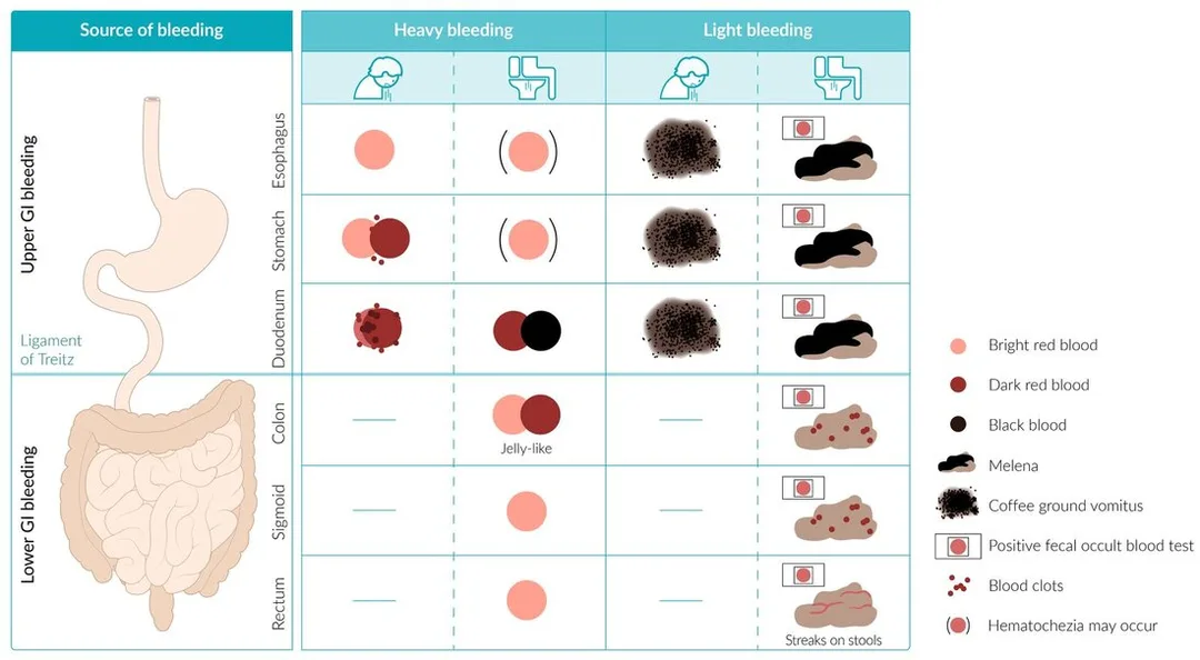
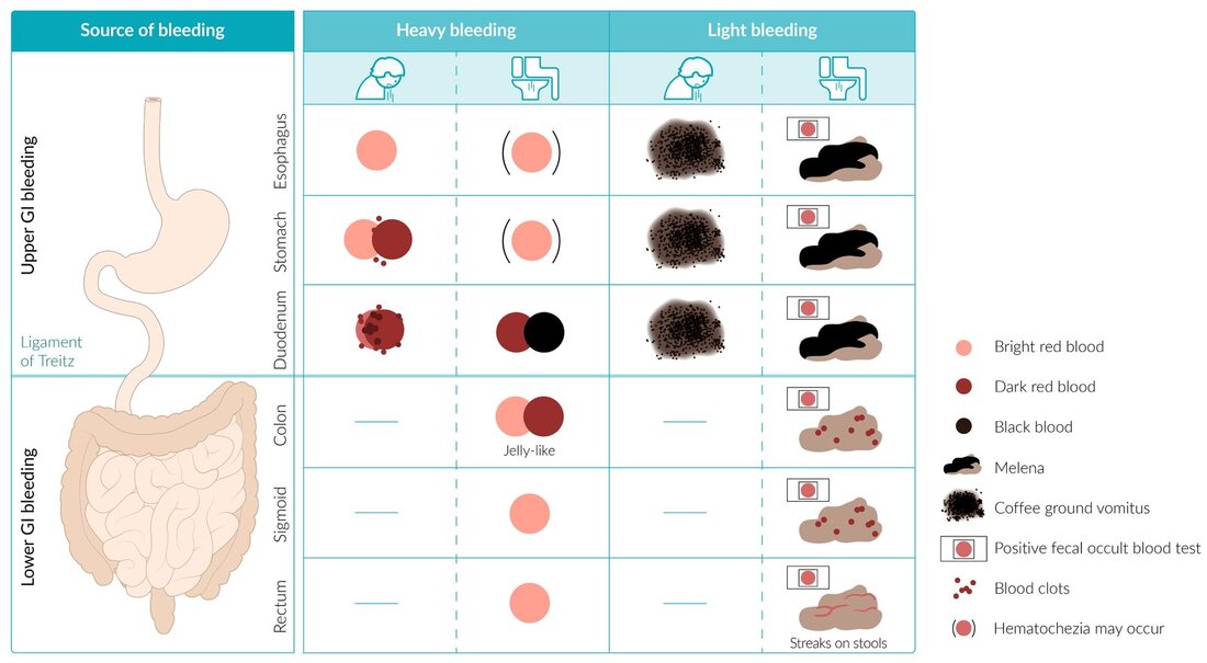
bleeding coming from the digestive tract's lower region, or from a location distal to the Treitz ligament.
Etiology
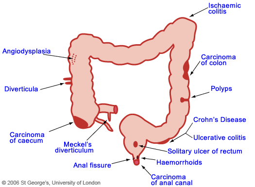
Colic polyps or carcinoma, anal fissures, haemorrhoids, diverticular disease and angiodysplasia (60–70%), and colitis (inflammatory, ulcerative, infectious, ischemic, radiation). Rarer conditions include endometriosis, small intestinal tumors, aorto-enteric fistula, Meckel's diverticulum, and a single rectal ulcer. Bleeding PR can occur in conjunction with brisk upper GI bleeding.
Epidemiology
Common, however less common than upper gastrointestinal bleeds (10–30% of GI bleeds).
more typical among older people.
H HISTORY
Fresh bleeding or bloody diarrhea suggests a hemorrhage that is distal to the caecum.
Cause-suggestive history: inflammatory bowel disease, for example.
Examination
indicators of chronic iron deficient anemia.
symptoms of shock, tachycardia, orthostatic or overt hypotension, and acute hypovolemia.
Investigation
Blood: FBC, U&Es, clotting, LFT, and blood crossmatching in extensive hemorrhages. The detection of occult blood loss is accomplished using fecal occult blood tests, or guaiac.
Endoscopy: Sigmoidoscopy/colonoscopy; blood will obstruct vision unless the bleeding rate is moderate. It can be utilized for colonic lesion diagnosis and therapy once the bleeding has stopped. OGD to rule out upper GI causes in severe, rapid bleeding.
Mesenteric angiography: Can identify the location of bleeding (sensitivity < 60%); however, to show the site, the bleeding rate needs to be more than 0.5 mL/minute and must occur during the contrast injection. The usage of CT and MR angiography is growing.
Scintigraphy: A radiolabelled 99mTc-RBC scan is less accurate in its particular localization and can detect bleeding at a rate of 0.1–0.35 mL/minute. Technetiumscan: for Meckel's diverticulum-related ectopic stomach mucosa.
Laparotomy and enteroscopy: Using an endoscope inserted through the gut wall, the whole colon and small bowel are examined for internal and exterior lesions.
Management
Sufficient IV access, proactive resuscitation, and coagulopathy correction include resuscitation.
OGD and NG tube search for an upper GI source. In cases of substantial bleeding, close observation in an ICU or HDU is necessary. The majority will accept cautious management.
Interventional radiology includes vasopressin infusion and angiography for bleeding vessel localization and transcatheter embolization.
Endoscopic: Lower GI endoscopy, such as laser photocoagulation of angiodysplasia, may also be used to treat the cause in stable individuals.
Surgical: A subtotal colectomy may be necessary in cases of severe or recurring bleeding, or if endoscopic or angiographic treatment is not feasible or has not worked.
Complications
anemia, shock from low blood volume, and collapse.
Prognosis
Depending on the reason, most will finish on their own. Diverticular illness is the most prevalent cause of potentially fatal lower gastrointestinal bleeding; yet, 90% of diverticular bleeding will resolve with conservative treatment. Early management and forceful resuscitation enhance the prognosis in cases of serious GI bleeding.
Introduction
bleeding coming from the digestive tract's lower region, or from a location distal to the Treitz ligament.
Etiology
Colic polyps or carcinoma, anal fissures, haemorrhoids, diverticular disease and angiodysplasia (60–70%), and colitis (inflammatory, ulcerative, infectious, ischemic, radiation). Rarer conditions include endometriosis, small intestinal tumors, aorto-enteric fistula, Meckel's diverticulum, and a single rectal ulcer. Bleeding PR can occur in conjunction with brisk upper GI bleeding.
Epidemiology
Common, however less common than upper gastrointestinal bleeds (10–30% of GI bleeds).
more typical among older people.
H HISTORY
Fresh bleeding or bloody diarrhea suggests a hemorrhage that is distal to the caecum.
Cause-suggestive history: inflammatory bowel disease, for example.
Examination
indicators of chronic iron deficient anemia.
symptoms of shock, tachycardia, orthostatic or overt hypotension, and acute hypovolemia.
Investigation
Blood: FBC, U&Es, clotting, LFT, and blood crossmatching in extensive hemorrhages. The detection of occult blood loss is accomplished using fecal occult blood tests, or guaiac.
Endoscopy: Sigmoidoscopy/colonoscopy; blood will obstruct vision unless the bleeding rate is moderate. It can be utilized for colonic lesion diagnosis and therapy once the bleeding has stopped. OGD to rule out upper GI causes in severe, rapid bleeding.
Mesenteric angiography: Can identify the location of bleeding (sensitivity < 60%); however, to show the site, the bleeding rate needs to be more than 0.5 mL/minute and must occur during the contrast injection. The usage of CT and MR angiography is growing.
Scintigraphy: A radiolabelled 99mTc-RBC scan is less accurate in its particular localization and can detect bleeding at a rate of 0.1–0.35 mL/minute. Technetiumscan: for Meckel's diverticulum-related ectopic stomach mucosa.
Laparotomy and enteroscopy: Using an endoscope inserted through the gut wall, the whole colon and small bowel are examined for internal and exterior lesions.
Management
Sufficient IV access, proactive resuscitation, and coagulopathy correction include resuscitation.
OGD and NG tube search for an upper GI source. In cases of substantial bleeding, close observation in an ICU or HDU is necessary. The majority will accept cautious management.
Interventional radiology includes vasopressin infusion and angiography for bleeding vessel localization and transcatheter embolization.
Endoscopic: Lower GI endoscopy, such as laser photocoagulation of angiodysplasia, may also be used to treat the cause in stable individuals.
Surgical: A subtotal colectomy may be necessary in cases of severe or recurring bleeding, or if endoscopic or angiographic treatment is not feasible or has not worked.
Complications
anemia, shock from low blood volume, and collapse.
Prognosis
Depending on the reason, most will finish on their own. Diverticular illness is the most prevalent cause of potentially fatal lower gastrointestinal bleeding; yet, 90% of diverticular bleeding will resolve with conservative treatment. Early management and forceful resuscitation enhance the prognosis in cases of serious GI bleeding.
0 Comments
