- Published on
Surgery - Lymphoedema
introduction
excessive buildup of lymphatic fluid in the extracellular space as a result of the lymphatic system's compromised performance.
Etiology
Primary (hereditary): The genetic alterations in the majority of families remain unknown.
Those named are:
Milroy's illness: A mutation in the vascular endothelial growth factor receptor VEGFR-3 gene results in autosomal dominant inheritance.
An inactivating mutation in the transcription factor FOXC2 gene causes lymphoedema distichiasis syndrome. correlated with additional eyelash rows.
Mutations in the transcription factor SOX18 gene cause the hypotrichosis-lymphoedema-telangiectasia syndrome.
Secondary (acquired): Occurs after blockage or damage of lymphatic channels, such as a TB infection, silica, or Wuchereria bancrofti infection (filariasis).
& regional lymphatics can be surgically excised or treated with post-radiotherapy for cancer, most often breast cancer.
malignant lymph node invasion in the area.
Epidemiology
Principal: Rare; yearly incidence 1/6000–10,000; 2-3 times more prevalent in females.
Secondary: Far more typical. The most frequent cause globally is filariasis.
History
can appear at any age (lymphoedema distichiasis), during puberty (Milroy's), or at birth. Lower limb swelling occurs most frequently gradually, getting worse towards the end of the day. Other times, the swelling is in the genitalia or arms. Chylothorax, chylous ascites, or leaky skin vesicles are possible presentations in megalymphatics.
Examination
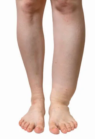
Skin oedema in its early stages is pitting. Subsequently, the skin turns brawny, fibrotic, and non-pitting; vesicles may start to cry. Toes get squared off, and ankles lose their shape. Areas of skin that become thicker (condylomas) may form.
Pathogenesis
Primary/congenital lymphoedema is caused by lymphatic valve hypoplasia, hyperplasia, or inadequate function. The buildup of protein-rich fluid and oedema that follows, frequently accompanied by inflammation, causes fibrosis and an overabundance of connective and adipose tissue, which causes swelling in the extremities.
Investigations
Isotope lymphography: a gamma counter is used to quantify movement after a subcutaneous injection of a colloid labeled with 99 mTc into the foot's first web space.
Contrast lymphangiography is no longer often used.
A CT or MRI scan can reveal lymphoedema-related abnormalities, such as oedema in the fascial planes or thickening of the skin. If an obstructive lesion is present, it can reveal the cause.
Other: tissue/lymph node biopsies or bioelectric impedance analysis.
Management
Conservative measures include massage using pneumatic devices or manual lymphatic drainage, limb elevation, exercise, graduated compression stockings, and skin or foot care. Venesection or blood pressure monitoring shouldn't be done on limbs that are impacted.
Medical: Infection treatment (e.g., cellulitis antibiotics, tinea pedis antifungals).
Diuretics don't have any advantages.
Surgery: Not often utilized. Surgical techniques employed comprise liposuction combined with forceful postoperative compression; the Homan's procedure entails generating skin flaps, removing subcutaneous tissue, and then resuturing the skin. After skin and subcutaneous tissue are removed, skin grafting is the next step in the Charles reduction process.
Complications
Boost Skin that is deficient in fluid and protein is more vulnerable to cellulitis, poor cosmesis, pain from enlarged tissue, decreased mobility, and ulceration.
Prognosis
Good, usually responds to conservative therapy for primary lymphoedema.
Depending on the cause, secondary lymphoedema may occur.
introduction
excessive buildup of lymphatic fluid in the extracellular space as a result of the lymphatic system's compromised performance.
Etiology
Primary (hereditary): The genetic alterations in the majority of families remain unknown.
Those named are:
Milroy's illness: A mutation in the vascular endothelial growth factor receptor VEGFR-3 gene results in autosomal dominant inheritance.
An inactivating mutation in the transcription factor FOXC2 gene causes lymphoedema distichiasis syndrome. correlated with additional eyelash rows.
Mutations in the transcription factor SOX18 gene cause the hypotrichosis-lymphoedema-telangiectasia syndrome.
Secondary (acquired): Occurs after blockage or damage of lymphatic channels, such as a TB infection, silica, or Wuchereria bancrofti infection (filariasis).
& regional lymphatics can be surgically excised or treated with post-radiotherapy for cancer, most often breast cancer.
malignant lymph node invasion in the area.
Epidemiology
Principal: Rare; yearly incidence 1/6000–10,000; 2-3 times more prevalent in females.
Secondary: Far more typical. The most frequent cause globally is filariasis.
History
can appear at any age (lymphoedema distichiasis), during puberty (Milroy's), or at birth. Lower limb swelling occurs most frequently gradually, getting worse towards the end of the day. Other times, the swelling is in the genitalia or arms. Chylothorax, chylous ascites, or leaky skin vesicles are possible presentations in megalymphatics.
Examination
Skin oedema in its early stages is pitting. Subsequently, the skin turns brawny, fibrotic, and non-pitting; vesicles may start to cry. Toes get squared off, and ankles lose their shape. Areas of skin that become thicker (condylomas) may form.
Pathogenesis
Primary/congenital lymphoedema is caused by lymphatic valve hypoplasia, hyperplasia, or inadequate function. The buildup of protein-rich fluid and oedema that follows, frequently accompanied by inflammation, causes fibrosis and an overabundance of connective and adipose tissue, which causes swelling in the extremities.
Investigations
Isotope lymphography: a gamma counter is used to quantify movement after a subcutaneous injection of a colloid labeled with 99 mTc into the foot's first web space.
Contrast lymphangiography is no longer often used.
A CT or MRI scan can reveal lymphoedema-related abnormalities, such as oedema in the fascial planes or thickening of the skin. If an obstructive lesion is present, it can reveal the cause.
Other: tissue/lymph node biopsies or bioelectric impedance analysis.
Management
Conservative measures include massage using pneumatic devices or manual lymphatic drainage, limb elevation, exercise, graduated compression stockings, and skin or foot care. Venesection or blood pressure monitoring shouldn't be done on limbs that are impacted.
Medical: Infection treatment (e.g., cellulitis antibiotics, tinea pedis antifungals).
Diuretics don't have any advantages.
Surgery: Not often utilized. Surgical techniques employed comprise liposuction combined with forceful postoperative compression; the Homan's procedure entails generating skin flaps, removing subcutaneous tissue, and then resuturing the skin. After skin and subcutaneous tissue are removed, skin grafting is the next step in the Charles reduction process.
Complications
Boost Skin that is deficient in fluid and protein is more vulnerable to cellulitis, poor cosmesis, pain from enlarged tissue, decreased mobility, and ulceration.
Prognosis
Good, usually responds to conservative therapy for primary lymphoedema.
Depending on the cause, secondary lymphoedema may occur.
0 Comments
