- Published on
Surgery - Malignant Melanoma
Introduction
malignancy brought on by the neoplastic alteration of skin pigment-forming melanocytes. the primary reason why skin diseases cause death. can also develop in the leptomeninges, GI tract, eye, or ear.
Etiology
Multiple factors contribute to the neoplastic transformation, with a build-up of genetic alterations. Inheritable mutations in the tumour suppressor genes CDKN2A and CDK4 have been found in 10% of cases of uncommon familial melanoma syndrome (autosomal dominant). Birth defects, such as xeroderma pigmentosum.
Four categories of histopathology:
(1) Superficial spreading (70%): Usually develops from an already-existing naevus, a flat or elevated brown lesion, potentially with variegate coloring, and it spreads radially prior to the vertical growth phase.
Nodular (15%): De novo, aggressive, ulcerative or bleed readily, lacks radial development phase.
Lentigo maligna (10%): This condition is more prevalent in elderly people who have sun exposure, has large, flat lesions, and grows more slowly. Typically on the arms, neck, or face.
(4) Acral lentiginous (5%): These lesions appear on the soles, palms, and subungual regions. most prevalent kind in non-White populations, with an often postponed diagnosis.
linked to UV radiation exposure (sun exposure, particularly if there is a history of blistering burns, tanning lamp use, and PUVA). Increased number of dysplastic moles and fair skin.
Epidemiology
steadily rising incidence: in the UK, 6,000 cases are diagnosed annually, whereas in the USA, the lifetime risk is 1/60. Non-white races are at a 20 percent risk from white races. causes 3/4 of skin cancer fatalities but just 4% of skin cancer cases.
H HISTORY
A pigmented skin lesion's size, form, or color changing, as well as any redness, bleeding, crusting, or ulceration. But more than 60% don't start from pre-existing moles.
Examination
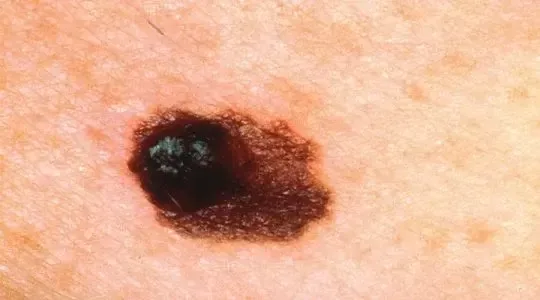
ABCDE criteria for examining moles:
A Asymmetry
B Border irregularity/bleeding
C Colour variation
D Diameter >6mm
E Elevation/evolving changes over time
Amelanotic melanoma is non-pigmented, often associated with nodular subtype or
metastases of undifferentiated melanoma to the skin.
Investigations
Excisional biopsy: To determine Breslow thickness or Clark's levels and provide a histological diagnosis.
Sentinel lymph node biopsy: Histological examination is used to determine the presence of metastases after lymphoscintigraphy and blue dye are used to identify the nodes.
Staging: CT, MRI, CXR, and PET scans for imaging.
Bloods: LFTs and LDH (metastases frequently occur in the liver).
Management
Primarily, prevent sunburn, limit excessive sun exposure, and raise public awareness.
Operation: Broad local excision with a margin that varies according to the depth of invasion (<1 mm: 1 cm, 1-4 mm: 2 cm border). Skin grafting can be necessary. If the melanoma is more than 1 mm, a sentinel lymph node biopsy is done; if the results are positive, a complete lymph node dissection is done. Careful follow-up is necessary after treatment.
Diseases with metastases: Chemotherapy: not very effective. Response to dacarbazine in combination with paclitaxel and cisplatin during clinical studies. ought to be included in a trial. High-dose interferon a-2b immunotherapy has been demonstrated in trials to enhance relapse-free survival, but not overall survival (for high-risk tumors >4mm with regional lymph node metastases). Research on vaccinations against melanoma is ongoing.
Complications
Localized: ulcers and bleeding.
Metastases: mass effect and bleeding.
Post-surgical: Block dissection of lymph nodes may cause wound issues or lymphoedema.
Prognosis
Five-year survival is 90–95% for lesions that are less than 1 mm deep, 13–69% for node-positive disease, and a 9-month median survival for metastatic disease. Every stage of cancer survival is decreased by ulceration.
Lesions in the trunk as opposed to the limb, ulceration, and an elevated mitotic rate are poorer prognostic indications.
The prognosis is worse for men than for women.
Introduction
malignancy brought on by the neoplastic alteration of skin pigment-forming melanocytes. the primary reason why skin diseases cause death. can also develop in the leptomeninges, GI tract, eye, or ear.
Etiology
Multiple factors contribute to the neoplastic transformation, with a build-up of genetic alterations. Inheritable mutations in the tumour suppressor genes CDKN2A and CDK4 have been found in 10% of cases of uncommon familial melanoma syndrome (autosomal dominant). Birth defects, such as xeroderma pigmentosum.
Four categories of histopathology:
(1) Superficial spreading (70%): Usually develops from an already-existing naevus, a flat or elevated brown lesion, potentially with variegate coloring, and it spreads radially prior to the vertical growth phase.
Nodular (15%): De novo, aggressive, ulcerative or bleed readily, lacks radial development phase.
Lentigo maligna (10%): This condition is more prevalent in elderly people who have sun exposure, has large, flat lesions, and grows more slowly. Typically on the arms, neck, or face.
(4) Acral lentiginous (5%): These lesions appear on the soles, palms, and subungual regions. most prevalent kind in non-White populations, with an often postponed diagnosis.
linked to UV radiation exposure (sun exposure, particularly if there is a history of blistering burns, tanning lamp use, and PUVA). Increased number of dysplastic moles and fair skin.
Epidemiology
steadily rising incidence: in the UK, 6,000 cases are diagnosed annually, whereas in the USA, the lifetime risk is 1/60. Non-white races are at a 20 percent risk from white races. causes 3/4 of skin cancer fatalities but just 4% of skin cancer cases.
H HISTORY
A pigmented skin lesion's size, form, or color changing, as well as any redness, bleeding, crusting, or ulceration. But more than 60% don't start from pre-existing moles.
Examination
ABCDE criteria for examining moles:
A Asymmetry
B Border irregularity/bleeding
C Colour variation
D Diameter >6mm
E Elevation/evolving changes over time
Amelanotic melanoma is non-pigmented, often associated with nodular subtype or
metastases of undifferentiated melanoma to the skin.
Investigations
Excisional biopsy: To determine Breslow thickness or Clark's levels and provide a histological diagnosis.
Sentinel lymph node biopsy: Histological examination is used to determine the presence of metastases after lymphoscintigraphy and blue dye are used to identify the nodes.
Staging: CT, MRI, CXR, and PET scans for imaging.
Bloods: LFTs and LDH (metastases frequently occur in the liver).
Management
Primarily, prevent sunburn, limit excessive sun exposure, and raise public awareness.
Operation: Broad local excision with a margin that varies according to the depth of invasion (<1 mm: 1 cm, 1-4 mm: 2 cm border). Skin grafting can be necessary. If the melanoma is more than 1 mm, a sentinel lymph node biopsy is done; if the results are positive, a complete lymph node dissection is done. Careful follow-up is necessary after treatment.
Diseases with metastases: Chemotherapy: not very effective. Response to dacarbazine in combination with paclitaxel and cisplatin during clinical studies. ought to be included in a trial. High-dose interferon a-2b immunotherapy has been demonstrated in trials to enhance relapse-free survival, but not overall survival (for high-risk tumors >4mm with regional lymph node metastases). Research on vaccinations against melanoma is ongoing.
Complications
Localized: ulcers and bleeding.
Metastases: mass effect and bleeding.
Post-surgical: Block dissection of lymph nodes may cause wound issues or lymphoedema.
Prognosis
Five-year survival is 90–95% for lesions that are less than 1 mm deep, 13–69% for node-positive disease, and a 9-month median survival for metastatic disease. Every stage of cancer survival is decreased by ulceration.
Lesions in the trunk as opposed to the limb, ulceration, and an elevated mitotic rate are poorer prognostic indications.
The prognosis is worse for men than for women.
0 Comments
