- Published on
Surgery - Prostate Carcinoma
Introduction
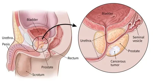
a primary prostate gland malignant tumor. 95% of the cases are adenocarcinomas, which vary in their level of differentiation.
Risk Factors
The largest risk factor is age. Afro-Caribbean people are more likely than Caucasian people to present with an aggressive disease at a younger age. Geographic distribution: lower in the Far East and higher in North America and Europe. Gene implicated on chromosome 1 in family history. Dietary factors: Consumption of meat, alcohol, and high fat foods is linked to reduced soy intake.
Epidemiology
The second leading cause of cancer-related mortality in men. 50–70 instances per 100,000 year.
80% of males over 80 years old who have autopsies had microfoci related to malignancy.
History
frequently asymptomatic and found through PSA testing.
Obstruction of the lower urinary tract: nocturia, terminal dribbling, weak stream, hesitation, and frequency.
Metastatic spread: Spinal cord compression or discomfort in the bones as a result of metastases.
Anorexia, weight loss, and malaise are common signs of cancer.
Examination
Asymmetrical hard nodular prostate gland with lack of the midline sulcus was seen during a digital rectal examination.
Investigations
Blood: bone profile, PSA, LFT, U&E, and FBC.
Antigen specific to prostates: Age-related and non-specific values may be higher in cases of prostatitis, benign prostatic hyperplasia, or after catheterization.
Transrectal ultrasonography (TRUS) and needle biopsy: The choice to proceed with TRUS and biopsy is guided by the PSA level and digital rectal examination. Different algorithms are used to make decisions, such as the following: -patients who, regardless of a digital rectal examination, have a PSA more than 10 ng/ml.
-individuals with free PSA <30% and a PSA of 4–10 ng/ml.
-individuals whose digital rectal examination revealed a palpable nodule.
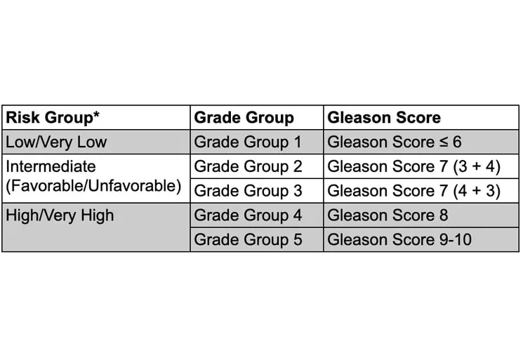
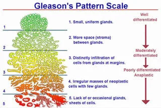
Gleason score: Two scores are assigned depending on predominant appearance, with a maximum score of 5 + 5 (10). Grading is based on histology from biopsy.
CT/MRI scan: Evaluates lymph node involvement and the degree of local invasion.
For bone metastases, use an isotope bone scan.
TNM staging scheme:
T1: confined to the prostate (a: one lobe; b: both lobes); T3: extending through capsule; T4: fixed tumor invading adjacent structures other than seminal vesicles; N1: regional lymph nodes involved; M: metastases. T1a: incidental <5% on TURP; T1b: incidental >5% on TURP; T1c: identified on needle biopsy.
Screening: In the United States, PSA values and digital rectal examination are employed. There is not enough evidence to support universal screening.
Management
Multidisciplinary discussion: on the best treatment option based on the patient's wishes, age, and comorbidities, as well as tumor staging.
Active monitoring For people with low and intermediate risks, watchful waiting along with PSA monitoring is usually the best course of action.
For tumors restricted to the gland, a radical prostatectomy may be necessary. Retropubic (for pelvic lymph node sample) or perineal approaches can be used for this. through laparoscopic, robotically assisted, or open surgery techniques.
Medical hormone therapy involves starting with an anti-androgen (cyproterone acetate) and LHRH analogues (goserelin, for example) to prevent a testosterone flare-up. Other treatments include anti-androgens, such as steroidal or nonsteroidal forms like flutamide and bicalutamide. is a neoadjuvant treatment option.
Surgery for androgen ablation: A bilateral orchidectomy can impede the course of the illness.
Chemotherapy: Hormone-refractory diseases may be treated with cetaxel.
Adjuvant radiation may be used in cases where the surgical resection margins are insufficient or where tumors that are limited to the pelvis affect lymph nodes. Moreover, brachytherapy is an option. It has been demonstrated that neoadjuvant hormone treatment works well for big but localized tumors before external beam radiation therapy. For the treatment of neurological issues and bone pain, palliative radiation therapy is an option.
Complications
Spread: Most frequently, blood-borne spread affects bone (particularly the spine), lung, and liver. Local growth occurs into seminal vesicles, the bladder, and the rectum. Lymphatic spread travels to the iliac and para-aortic nodes.
From disease: metastatic illness, hypercalcemia, obstructive hydronephrosis.
Following surgery: urethral stricture, impotence, and incontinence.
bladder and bowel damage from radiation.
Androgen shortage can result in libido loss, gynecomastia, hot flashes, impotence, and osteoporosis through hormone therapy. Anti-androgen treatment resistance due to tumour hormone escape.
Prognosis
Radical therapy yields a 10-year survival of more than 80%; untreated patients had an 80% 5-year survival.
The median survival time for metastatic illness is 18–24 months.
Introduction
a primary prostate gland malignant tumor. 95% of the cases are adenocarcinomas, which vary in their level of differentiation.
Risk Factors
The largest risk factor is age. Afro-Caribbean people are more likely than Caucasian people to present with an aggressive disease at a younger age. Geographic distribution: lower in the Far East and higher in North America and Europe. Gene implicated on chromosome 1 in family history. Dietary factors: Consumption of meat, alcohol, and high fat foods is linked to reduced soy intake.
Epidemiology
The second leading cause of cancer-related mortality in men. 50–70 instances per 100,000 year.
80% of males over 80 years old who have autopsies had microfoci related to malignancy.
History
frequently asymptomatic and found through PSA testing.
Obstruction of the lower urinary tract: nocturia, terminal dribbling, weak stream, hesitation, and frequency.
Metastatic spread: Spinal cord compression or discomfort in the bones as a result of metastases.
Anorexia, weight loss, and malaise are common signs of cancer.
Examination
Asymmetrical hard nodular prostate gland with lack of the midline sulcus was seen during a digital rectal examination.
Investigations
Blood: bone profile, PSA, LFT, U&E, and FBC.
Antigen specific to prostates: Age-related and non-specific values may be higher in cases of prostatitis, benign prostatic hyperplasia, or after catheterization.
Transrectal ultrasonography (TRUS) and needle biopsy: The choice to proceed with TRUS and biopsy is guided by the PSA level and digital rectal examination. Different algorithms are used to make decisions, such as the following: -patients who, regardless of a digital rectal examination, have a PSA more than 10 ng/ml.
-individuals with free PSA <30% and a PSA of 4–10 ng/ml.
-individuals whose digital rectal examination revealed a palpable nodule.
Gleason score: Two scores are assigned depending on predominant appearance, with a maximum score of 5 + 5 (10). Grading is based on histology from biopsy.
CT/MRI scan: Evaluates lymph node involvement and the degree of local invasion.
For bone metastases, use an isotope bone scan.
TNM staging scheme:
T1: confined to the prostate (a: one lobe; b: both lobes); T3: extending through capsule; T4: fixed tumor invading adjacent structures other than seminal vesicles; N1: regional lymph nodes involved; M: metastases. T1a: incidental <5% on TURP; T1b: incidental >5% on TURP; T1c: identified on needle biopsy.
Screening: In the United States, PSA values and digital rectal examination are employed. There is not enough evidence to support universal screening.
Management
Multidisciplinary discussion: on the best treatment option based on the patient's wishes, age, and comorbidities, as well as tumor staging.
Active monitoring For people with low and intermediate risks, watchful waiting along with PSA monitoring is usually the best course of action.
For tumors restricted to the gland, a radical prostatectomy may be necessary. Retropubic (for pelvic lymph node sample) or perineal approaches can be used for this. through laparoscopic, robotically assisted, or open surgery techniques.
Medical hormone therapy involves starting with an anti-androgen (cyproterone acetate) and LHRH analogues (goserelin, for example) to prevent a testosterone flare-up. Other treatments include anti-androgens, such as steroidal or nonsteroidal forms like flutamide and bicalutamide. is a neoadjuvant treatment option.
Surgery for androgen ablation: A bilateral orchidectomy can impede the course of the illness.
Chemotherapy: Hormone-refractory diseases may be treated with cetaxel.
Adjuvant radiation may be used in cases where the surgical resection margins are insufficient or where tumors that are limited to the pelvis affect lymph nodes. Moreover, brachytherapy is an option. It has been demonstrated that neoadjuvant hormone treatment works well for big but localized tumors before external beam radiation therapy. For the treatment of neurological issues and bone pain, palliative radiation therapy is an option.
Complications
Spread: Most frequently, blood-borne spread affects bone (particularly the spine), lung, and liver. Local growth occurs into seminal vesicles, the bladder, and the rectum. Lymphatic spread travels to the iliac and para-aortic nodes.
From disease: metastatic illness, hypercalcemia, obstructive hydronephrosis.
Following surgery: urethral stricture, impotence, and incontinence.
bladder and bowel damage from radiation.
Androgen shortage can result in libido loss, gynecomastia, hot flashes, impotence, and osteoporosis through hormone therapy. Anti-androgen treatment resistance due to tumour hormone escape.
Prognosis
Radical therapy yields a 10-year survival of more than 80%; untreated patients had an 80% 5-year survival.
The median survival time for metastatic illness is 18–24 months.
0 Comments
