- Published on
Surgery - Renal Carcinoma
Introduction
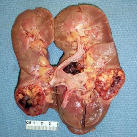
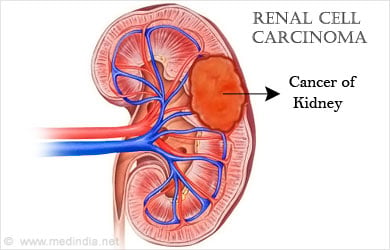
Malignancy originating from the renal tubular epithelium.
Etiology
Mutations in the oncogene (MET) or tumor suppressor genes (VHL, TSC) are linked to sporadic and hereditary forms.
Von Hippel-Lindau disease, hereditary papillary renal carcinoma, familial renal oncocytoma, and hereditary renal carcinoma are examples of hereditary syndromes.
Risk Factors
Chronic dialysis is used to treat acquired cystic disease of the kidney, obesity, smoking, and tuberous sclerosis.
Epidemiology
Rare (three percent of adult cancer cases). 3:1 male to female. grows with age; in men over 60, 75% of cases occur.
History
90% of the time asymptomatic, accidental discovery made when scanning. the traditional trio of abdominal mass, flank pain, and haematuria (only 10% of patients).
Systemic indications of cancer: Loss of weight, lethargy, and paraneoplastic disorders, such as polycythaemia or hypercalcaemia-related pyrexia, are also present.
Examination
Anaemia, plethora, hypertension, or no symptoms could exist. renal tumor that is palpable.A left-sided varicocoele can result from occlusion of the left testicular vein by a left-sided tumor that extends into the left renal vein. In terms of presentation, one-third had metastases.
Investigations
Urine: Cytology, Dipstick (for haematuria detection).
FBC, U&Es, Ca2+, LFTs (linked to paraneoplastic syndromes such as polycythemia, hypercalcemia, abnormal LFTs in the absence of liver metastases, and Stauffer syndrome), and ESR in 75% of the blood samples.
Imaging: bone and PET scanning for metastases; ultrasound, CT, or MRI scans.
Pathology: Clear cell, chromophilic, chromophobic, oncocytoma, and collecting duct are among the histological forms.
Robson staging : I: tumor inside the renal capsule. II: invades the perinephric fat but not Gerota's fascia . III: Invades local lymph nodes, the IVC, or the renal vein. IV: infiltrates distant metastases or nearby viscera.
Management
Surgery : The usual procedure is a radical nephrectomy, which involves the removal of the ipsilateral adrenal gland, regional lymphadenectomy, Gerota's fascia, and perinephric fat. can be carried out using laparoscopic, thoraco-abdominal, transperitoneal, or flank methods.
Radiation therapy and chemotherapy: Renal Carcinoma is well known for its chemotherapeutic agent resistance, which is mediated by p-glycoprotein, a multidrug resistance protein. With more recent multikinase inhibitors like sunitinib and sorafenib, positive results are achievable. Metastatic lesions may be treated with radiation therapy.
Complications
50% of distant metastases impact the lung, and 33% the bone. Local invasion, such as the blockage of the IVC or the invasion of perinephric fat. Localized bleeding and clot colic. Paraneoplastic Syndrome.
Prognosis
Depending on the tumor's nature and stage. Five-year survival after stage I disease excision is 94%; nodal spread accounts for 18–30% of the survival, whereas distant metastases account for 0–20%.
Introduction
Malignancy originating from the renal tubular epithelium.
Etiology
Mutations in the oncogene (MET) or tumor suppressor genes (VHL, TSC) are linked to sporadic and hereditary forms.
Von Hippel-Lindau disease, hereditary papillary renal carcinoma, familial renal oncocytoma, and hereditary renal carcinoma are examples of hereditary syndromes.
Risk Factors
Chronic dialysis is used to treat acquired cystic disease of the kidney, obesity, smoking, and tuberous sclerosis.
Epidemiology
Rare (three percent of adult cancer cases). 3:1 male to female. grows with age; in men over 60, 75% of cases occur.
History
90% of the time asymptomatic, accidental discovery made when scanning. the traditional trio of abdominal mass, flank pain, and haematuria (only 10% of patients).
Systemic indications of cancer: Loss of weight, lethargy, and paraneoplastic disorders, such as polycythaemia or hypercalcaemia-related pyrexia, are also present.
Examination
Anaemia, plethora, hypertension, or no symptoms could exist. renal tumor that is palpable.A left-sided varicocoele can result from occlusion of the left testicular vein by a left-sided tumor that extends into the left renal vein. In terms of presentation, one-third had metastases.
Investigations
Urine: Cytology, Dipstick (for haematuria detection).
FBC, U&Es, Ca2+, LFTs (linked to paraneoplastic syndromes such as polycythemia, hypercalcemia, abnormal LFTs in the absence of liver metastases, and Stauffer syndrome), and ESR in 75% of the blood samples.
Imaging: bone and PET scanning for metastases; ultrasound, CT, or MRI scans.
Pathology: Clear cell, chromophilic, chromophobic, oncocytoma, and collecting duct are among the histological forms.
Robson staging : I: tumor inside the renal capsule. II: invades the perinephric fat but not Gerota's fascia . III: Invades local lymph nodes, the IVC, or the renal vein. IV: infiltrates distant metastases or nearby viscera.
Management
Surgery : The usual procedure is a radical nephrectomy, which involves the removal of the ipsilateral adrenal gland, regional lymphadenectomy, Gerota's fascia, and perinephric fat. can be carried out using laparoscopic, thoraco-abdominal, transperitoneal, or flank methods.
Radiation therapy and chemotherapy: Renal Carcinoma is well known for its chemotherapeutic agent resistance, which is mediated by p-glycoprotein, a multidrug resistance protein. With more recent multikinase inhibitors like sunitinib and sorafenib, positive results are achievable. Metastatic lesions may be treated with radiation therapy.
Complications
50% of distant metastases impact the lung, and 33% the bone. Local invasion, such as the blockage of the IVC or the invasion of perinephric fat. Localized bleeding and clot colic. Paraneoplastic Syndrome.
Prognosis
Depending on the tumor's nature and stage. Five-year survival after stage I disease excision is 94%; nodal spread accounts for 18–30% of the survival, whereas distant metastases account for 0–20%.
0 Comments
