- Published on
Surgery - Repair of an Abdominal Aortic Aneurysm (Open)
Introduction
Large asymptomatic aneurysms (diameter greater than 5.5 cm) are elective.
Aneurysms that are growing (>0.5 cm in 1 year).
Aneurysms that leak or burst are emergencies. Manifest aneurysms.
Anatomy
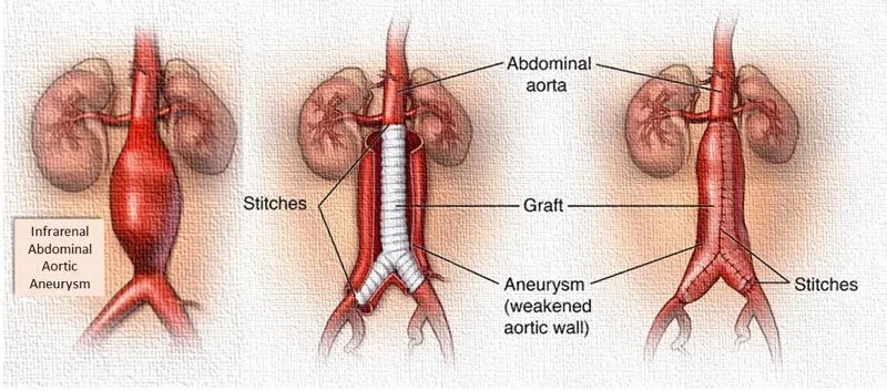
An aberrant focal dilatation of the abdominal aorta is known as an abdominal aortic aneurysm. Ninety-five percent start below the origin of the renal artery and may spread to the iliac arteries.
Usually fusiform in shape, their rate of expansion ranges from 0.2 to 0.8 cm/year, and the diameter determines the risk of rupture. Within the aneurysm, laminated thrombus builds up and may embolise distantly.
Investigations
Ultrasound and CT/MRI scanning are used to evaluate the size and anatomy of aneurysms during elective repairs. Reconstruction from 3D CT enables endovascular planning.
Pre-operative tests include FBC, coagulation, U&Es, crossmatch (six to eight units of blood), echocardiography, CXR, ECG, and cardiopulmonary.
After surgery: Monitor HDU and ITU settings closely. Check for emboli in the lower limbs. DVT prevention.
Actions
Access: To improve epidural pain control, a transverse incision or a full-length midline laparotomy are also options. rarely carried out as laparoscopic surgery with manual assistance.
Exposure: To avoid damaging the left sympathetic chain, the small bowel is moved upward and to the right, exposing the retroperitoneum over the aorta, which is slightly incised to the right. With caution to prevent harm to the left renal vein, which passes in front of the aorta, dissection is performed to expose the aorta from the infrarenal aorta to the bifurcation. It is determined, split, and ligated to identify the inferior mesenteric artery.
The aneurysm's proximal and distal ends are clamped, and systemic heparin is given.
Aneurysm opening: The contents of the aneurysm are visible when the aneurysm is opened longitudinally.
Sutures are used to limit any bleeding from the lumbar arteries in the back wall and to remove any thrombus inside the aneurysm.
Graft insertion: The distal aortic and bifurcation walls are examined. A tube graft is used to treat an aortic aneurysm. An aorto-iliac or, less frequently, an aorto-bifemoral trouser graft is utilized when the illness affects the proximal or distal iliacs. Prolene sutures are used to secure the grafts in position, and air or debris is flushed out afterwards.
Evaluation of the graft: The distal end is progressively opened with close supervision because of the possibility of hypotension and arrhythmias, after the aortic clamp is gradually relaxed to ensure hemostasis. After that, the aneurysm sac is sealed around the graft and sutured using the anastomosis suture line to prevent adhesions.
Closure: Using sutures or clips to close the skin in bulk.
When an aneurysm ruptures unexpectedly, the patient is taken to the operating room while having a systolic blood pressure of 80–100 mmHg. The procedure begins as soon as the patient is quickly draped and readied, with a "crash induction" of anesthesia. The bleeding vessel is to be rapidly clamped down and controlled.
Complications
bleeding, myocardial ischemia, MI or arrhythmias, cerebrovascular accident, respiratory problems (ARDS, infection, and atherosis), ischemia of the colon, ischemia of the spine, atheromatous embolization, renal failure, graft thrombosis, and endoleak. Late: False aneurysm at anastomosis, aorto-enteric fistula, and graft infection.
Prognosis
In most facilities, the rate of death from elective surgery is currently less than 5%. The mortality rate is exceedingly high when an aortic aneurysm ruptures or leaks and needs emergency repair.
0 Comments
