- Published on
Surgery - Skin cancer (basal cell carcinoma)
Introduction
Most prevalent type of skin cancer; also referred to as a "rodent ulcer"
Etiology
prolonged exposure to UV light or the sun. correlated with anomalies of the hedgehog/patched intracellular signaling cascade, as observed in naevoid basal cell carcinoma syndrome, or Gorlin's syndrome. Pitch that becomes photosensitized, tar, and arsenic are additional danger concerns.
Epidemiology
Frequent in the elderly, uncommon before the age of 40, common in people with fair skin and locations with considerable sun exposure. For Caucasians, the lifetime risk is 1:3.
History
a persistent, gradually developing skin lesion that mainly affects the face but can also affect the scalp, ears, or trunk.
Examination
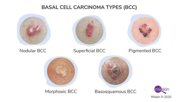
Most frequently, nodulo-ulcerative: tiny, shimmering, translucent skin covering a colored papule that initially grows slowly or a core ulcer known as a "rodent ulcer" that has elevated, pearly margins. The surface of the tumor is commonly covered in fine telangiectatic arteries. Lesions that are bigger and more protuberant may exhibit cystic transformation.
Morphoeic: Waxy, yellow-white, expanding plaque with a fuzzy border that tends to be more aggressive.
Superficial: Usually on the trunk, several brown or pink scaly plaques with a thin "whipcord" border that steadily expand to a diameter of more than 10 cm.
Pigmented: Any kind of basal cell carcinoma may have small amounts of brown or black pigment.
Pathogenesis
The dermis is invaded by small, dark blue-staining basal cells that form in distinct clusters and have palisade-like arrangements on their outer layer. There are several mitotic and apoptotic entities visible. Typically, growth is subtle, consistent, and sluggish. Although it doesn't spread, it has the ability to infiltrate and kill nearby tissues.
Investigations
Rarely is a biopsy required (clinical suspicion is the primary basis for diagnosis).
Management
For minor superficial lesions, photodynamic treatment, curettage, cauterization, and cryotherapy are employed.
Operative: For discrete nodular or cystic nodules, excision is recommended with a 0.5 cm margin of surrounding normal skin; for large tumors (1 cm in diameter) and lesions close to the eyes, nose, and ears, Mohs' micrographic surgery is the preferred method of treatment. This procedure involves a meticulous examination of tissue removed under frozen section. There may be a need for excision and skin flap covering.
Radiation therapy: beneficial for basal cell carcinomas affecting tissues (such as eyelids and tearducts) that are challenging to repair surgically. There is a chance of adverse effects include radiation dermatitis, ulceration, and depilation, and repeated treatments might be required.
Complications
The tumor progresses slowly but unabatedly. can cause facial disfigurement. has the ability to infiltrate and cause blindness in the orbit.
Prognosis
good when given the proper care. If ignored, it could spread, infiltrate, and become ulcerated. To identify further lesions or a local recurrence, routine follow-up is required.
Introduction
Most prevalent type of skin cancer; also referred to as a "rodent ulcer"
Etiology
prolonged exposure to UV light or the sun. correlated with anomalies of the hedgehog/patched intracellular signaling cascade, as observed in naevoid basal cell carcinoma syndrome, or Gorlin's syndrome. Pitch that becomes photosensitized, tar, and arsenic are additional danger concerns.
Epidemiology
Frequent in the elderly, uncommon before the age of 40, common in people with fair skin and locations with considerable sun exposure. For Caucasians, the lifetime risk is 1:3.
History
a persistent, gradually developing skin lesion that mainly affects the face but can also affect the scalp, ears, or trunk.
Examination
Most frequently, nodulo-ulcerative: tiny, shimmering, translucent skin covering a colored papule that initially grows slowly or a core ulcer known as a "rodent ulcer" that has elevated, pearly margins. The surface of the tumor is commonly covered in fine telangiectatic arteries. Lesions that are bigger and more protuberant may exhibit cystic transformation.
Morphoeic: Waxy, yellow-white, expanding plaque with a fuzzy border that tends to be more aggressive.
Superficial: Usually on the trunk, several brown or pink scaly plaques with a thin "whipcord" border that steadily expand to a diameter of more than 10 cm.
Pigmented: Any kind of basal cell carcinoma may have small amounts of brown or black pigment.
Pathogenesis
The dermis is invaded by small, dark blue-staining basal cells that form in distinct clusters and have palisade-like arrangements on their outer layer. There are several mitotic and apoptotic entities visible. Typically, growth is subtle, consistent, and sluggish. Although it doesn't spread, it has the ability to infiltrate and kill nearby tissues.
Investigations
Rarely is a biopsy required (clinical suspicion is the primary basis for diagnosis).
Management
For minor superficial lesions, photodynamic treatment, curettage, cauterization, and cryotherapy are employed.
Operative: For discrete nodular or cystic nodules, excision is recommended with a 0.5 cm margin of surrounding normal skin; for large tumors (1 cm in diameter) and lesions close to the eyes, nose, and ears, Mohs' micrographic surgery is the preferred method of treatment. This procedure involves a meticulous examination of tissue removed under frozen section. There may be a need for excision and skin flap covering.
Radiation therapy: beneficial for basal cell carcinomas affecting tissues (such as eyelids and tearducts) that are challenging to repair surgically. There is a chance of adverse effects include radiation dermatitis, ulceration, and depilation, and repeated treatments might be required.
Complications
The tumor progresses slowly but unabatedly. can cause facial disfigurement. has the ability to infiltrate and cause blindness in the orbit.
Prognosis
good when given the proper care. If ignored, it could spread, infiltrate, and become ulcerated. To identify further lesions or a local recurrence, routine follow-up is required.
0 Comments
