- Published on
Surgery - Skin Grafts and Flaps
Indications
Wounds where secondary intention healing or primary closure is not possible, would be deformative, or take a lengthy period, such as burns with traumatic skin loss, pressure sores, ulcers, or post-wide excision following tumor treatment or infection, like necrotizing fasciitis.
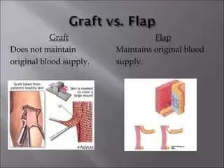
Skin grafts: A skin graft involves transferring a portion of skin from a donor site to a recipient site.
They need to be revascularized from the recipient location because they are free grafts. There are two types of skin grafts: split thickness (Thiersch graft) and full thickness (Wolfe graft). Split skin grafts can have poor visual appearance, a tendency to contract, and increased susceptibility to injury. Full thickness grafts have less contraction, more resilience, and a more appealing appearance. However, they are less dependable than split grafts and can sometimes only cover a small region.
Blocks of tissue that carry their own blood supply are known as flaps. The blood supply and movement of flaps, such as advancement or free flaps (the latter involving vascular or microvascular anastomosis), as well as the tissue content, which can be single (cutaneous, fascial, or bone) or composite (myocutaneous or fasciocutaneous), can all be used to classify flaps.
Flaps have the ability to correct a wide range of abnormalities and typically heal more quickly than grafts. The degree of experience needed and the possibility of functional or esthetic flaws remaining at the donor site are drawbacks.
Anatomy
Skin grafts with partial thickness comprise both the epidermis and the outermost layer of the dermis.
The deeper regions of sweat glands and hair follicles are where epidermis regenerates. Grafts can be meshed in order to cover a larger area and allow serous or serosanguinous fluid to escape, giving the appearance of a "string vest." Extra skin can be kept in the refrigerator aseptically for up to eight days, ideally for usage.
The dermis and epidermis are present in full thickness skin grafts.Postauricular, supraclavicular, lateral groin crease, and medial arm are common donor sites. They are limited in size, and donor sites are usually closed or may even need to have split skin grafted.
Investigational studies
Pre-op: Cleansing the recipient's wound and using a swab to make sure there are no infections. As they can inhibit graft taking, Streptococcus pyogenes and Pseudomonas should be treated initially. If anemia is substantial, treat it.
Observant wound care following surgery. The graft must not move while being bandaged or subsequently, due to the importance of dressings, to avoid the danger of failure. evaluation of viability or infection-related symptoms.
Actions
a wide range of approaches based on the donor and recipient locations.
Use a Humby knife to perform split skin transplants. Thighs and buttocks are common places for donors. Grafts are fastened at the margins using glue, staples, or sutures. Granulation tissue is the ideal location for skin grafts; other locations need to have adequate blood flow; naked tendon, cartilage, or bone cannot support a skin graft but can be covered by a skin flap.
After the skin transplant is placed, it is carefully dressed to help prevent shearing, infection, and hemorrhage or seroma.
Skin grafts done in their entirety: To aid in closure, the defect's pattern is noted on the donor site and an ellipse is carved around the graft. After that, the graft is properly prepared and sutured to the recipient site without causing any stress.
Skin flaps: A block of skin is transferred from a donor site to a recipient site, taking with it the blood supply of the underlying tissue (such as fascia, muscle, or bone). A local flap can be moved using a variety of methods, such as advancement, rotation, and transposition.
Complications
hemorrhage, infection, necrosis of the flap, scarring, contraction, and inadequate cosmesis.
Indications
Wounds where secondary intention healing or primary closure is not possible, would be deformative, or take a lengthy period, such as burns with traumatic skin loss, pressure sores, ulcers, or post-wide excision following tumor treatment or infection, like necrotizing fasciitis.
Skin grafts: A skin graft involves transferring a portion of skin from a donor site to a recipient site.
They need to be revascularized from the recipient location because they are free grafts. There are two types of skin grafts: split thickness (Thiersch graft) and full thickness (Wolfe graft). Split skin grafts can have poor visual appearance, a tendency to contract, and increased susceptibility to injury. Full thickness grafts have less contraction, more resilience, and a more appealing appearance. However, they are less dependable than split grafts and can sometimes only cover a small region.
Blocks of tissue that carry their own blood supply are known as flaps. The blood supply and movement of flaps, such as advancement or free flaps (the latter involving vascular or microvascular anastomosis), as well as the tissue content, which can be single (cutaneous, fascial, or bone) or composite (myocutaneous or fasciocutaneous), can all be used to classify flaps.
Flaps have the ability to correct a wide range of abnormalities and typically heal more quickly than grafts. The degree of experience needed and the possibility of functional or esthetic flaws remaining at the donor site are drawbacks.
Anatomy
Skin grafts with partial thickness comprise both the epidermis and the outermost layer of the dermis.
The deeper regions of sweat glands and hair follicles are where epidermis regenerates. Grafts can be meshed in order to cover a larger area and allow serous or serosanguinous fluid to escape, giving the appearance of a "string vest." Extra skin can be kept in the refrigerator aseptically for up to eight days, ideally for usage.
The dermis and epidermis are present in full thickness skin grafts.Postauricular, supraclavicular, lateral groin crease, and medial arm are common donor sites. They are limited in size, and donor sites are usually closed or may even need to have split skin grafted.
Investigational studies
Pre-op: Cleansing the recipient's wound and using a swab to make sure there are no infections. As they can inhibit graft taking, Streptococcus pyogenes and Pseudomonas should be treated initially. If anemia is substantial, treat it.
Observant wound care following surgery. The graft must not move while being bandaged or subsequently, due to the importance of dressings, to avoid the danger of failure. evaluation of viability or infection-related symptoms.
Actions
a wide range of approaches based on the donor and recipient locations.
Use a Humby knife to perform split skin transplants. Thighs and buttocks are common places for donors. Grafts are fastened at the margins using glue, staples, or sutures. Granulation tissue is the ideal location for skin grafts; other locations need to have adequate blood flow; naked tendon, cartilage, or bone cannot support a skin graft but can be covered by a skin flap.
After the skin transplant is placed, it is carefully dressed to help prevent shearing, infection, and hemorrhage or seroma.
Skin grafts done in their entirety: To aid in closure, the defect's pattern is noted on the donor site and an ellipse is carved around the graft. After that, the graft is properly prepared and sutured to the recipient site without causing any stress.
Skin flaps: A block of skin is transferred from a donor site to a recipient site, taking with it the blood supply of the underlying tissue (such as fascia, muscle, or bone). A local flap can be moved using a variety of methods, such as advancement, rotation, and transposition.
Complications
hemorrhage, infection, necrosis of the flap, scarring, contraction, and inadequate cosmesis.
0 Comments
