- Published on
Surgery - Tracheostomy
Indications
Longer-term ventilatory support (or weaning) is the most prevalent indication; additional conditions include head and neck surgery, neurological impairment (such as coma, stroke, and motor neurone disease), trauma, and bilateral vocal cord paralysis.
Anatomy
The posterior (membranous) portion of the trachea is composed of incomplete semicircular cartilage rings. The front surface of the trachea is convex and is covered (superiorly to inferiorly) by the cervical fascia, the inferior thyroid veins, the thyroid gland's isthmus (over the second and third rings), and the neck's strap muscles. The inferior thyroid veins and recurrent laryngeal nerves are located laterally in the tracheooesophageal grooves and are susceptible to damage.
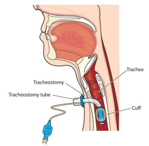
A tracheostomy can be cuffed or uncuffed, made of plastic, silicone, or metal, or fenestrated, which permits speech since the tube has a hole that lets air pass through the upper airway in the event that the external opening is blocked. Most have an obdurator that facilitates insertion but is removed once in place, an exterior cannula, and an interior cannula.
Investigations
carried out while under general anesthesia.
Pre-procedure: Pre-procedure counseling regarding risks and benefits is advised whenever it is feasible.
After surgery, meticulous attention is necessary. Regular sterile suction of retained secretions along with humidified gases. Positive pressure breathing is possible with cuffed tracheal tubes, although pressure necrosis can be avoided by routinely monitoring and deflating the tube. The tracheostomy tube can be changed after 5-7 days.
Actions
Access: The patient's neck is stretched in this position. In the middle of the cricoid cartilage and the sternal notch, a vertical or transverse incision is made. In order to reveal the trachea, cut through the platysma and subcutaneous tissue, split and retract the strap muscles, and separate the thyroid isthmus if required.
Surgical tracheostomy: Cut the trachea longitudinally or in a U-shape, creating two to three tracheal rings. In order to facilitate replacement in the event that the tracheostomy tube becomes dislodged, stay sutures can be positioned laterally to aid in maintaining the trachea open.
Put in a tracheostomy tube (removing the endotracheal tube at the same time might be required). The tracheostomy tube's balloon should be inflated before being sutured to the skin.
In ITU/HDU settings, percutaneous tracheostomy can be carried out. uses a bronchoscope and the Seldinger technique to create a percutaneous tracheal cannulation. A guidewire and progressive dilators are then inserted before the tracheostomy tube is inserted. Patients with complex anatomy, obesity, coagulopathy, or thyroid enlargement should not use this treatment.
Complications
Short-term: bleeding, infection, subcutaneous emphysema, pneumothorax or pneumomediastinum, damage to surrounding structures, such as oesophagus, arteries, recurrent laryngeal nerves, tube displacement, tracheal ulcers, tracheitis, mucus plugging, aspiration.
Prolonged: tracheomalacia, overgranulation, tracheo-oesophageal fistula (<1%), tracheocutaneous fistula, tracheal stenosis.
Indications
Longer-term ventilatory support (or weaning) is the most prevalent indication; additional conditions include head and neck surgery, neurological impairment (such as coma, stroke, and motor neurone disease), trauma, and bilateral vocal cord paralysis.
Anatomy
The posterior (membranous) portion of the trachea is composed of incomplete semicircular cartilage rings. The front surface of the trachea is convex and is covered (superiorly to inferiorly) by the cervical fascia, the inferior thyroid veins, the thyroid gland's isthmus (over the second and third rings), and the neck's strap muscles. The inferior thyroid veins and recurrent laryngeal nerves are located laterally in the tracheooesophageal grooves and are susceptible to damage.
A tracheostomy can be cuffed or uncuffed, made of plastic, silicone, or metal, or fenestrated, which permits speech since the tube has a hole that lets air pass through the upper airway in the event that the external opening is blocked. Most have an obdurator that facilitates insertion but is removed once in place, an exterior cannula, and an interior cannula.
Investigations
carried out while under general anesthesia.
Pre-procedure: Pre-procedure counseling regarding risks and benefits is advised whenever it is feasible.
After surgery, meticulous attention is necessary. Regular sterile suction of retained secretions along with humidified gases. Positive pressure breathing is possible with cuffed tracheal tubes, although pressure necrosis can be avoided by routinely monitoring and deflating the tube. The tracheostomy tube can be changed after 5-7 days.
Actions
Access: The patient's neck is stretched in this position. In the middle of the cricoid cartilage and the sternal notch, a vertical or transverse incision is made. In order to reveal the trachea, cut through the platysma and subcutaneous tissue, split and retract the strap muscles, and separate the thyroid isthmus if required.
Surgical tracheostomy: Cut the trachea longitudinally or in a U-shape, creating two to three tracheal rings. In order to facilitate replacement in the event that the tracheostomy tube becomes dislodged, stay sutures can be positioned laterally to aid in maintaining the trachea open.
Put in a tracheostomy tube (removing the endotracheal tube at the same time might be required). The tracheostomy tube's balloon should be inflated before being sutured to the skin.
In ITU/HDU settings, percutaneous tracheostomy can be carried out. uses a bronchoscope and the Seldinger technique to create a percutaneous tracheal cannulation. A guidewire and progressive dilators are then inserted before the tracheostomy tube is inserted. Patients with complex anatomy, obesity, coagulopathy, or thyroid enlargement should not use this treatment.
Complications
Short-term: bleeding, infection, subcutaneous emphysema, pneumothorax or pneumomediastinum, damage to surrounding structures, such as oesophagus, arteries, recurrent laryngeal nerves, tube displacement, tracheal ulcers, tracheitis, mucus plugging, aspiration.
Prolonged: tracheomalacia, overgranulation, tracheo-oesophageal fistula (<1%), tracheocutaneous fistula, tracheal stenosis.
0 Comments
