- Published on
Surgery - Urinary Tract Calculi
Definition
stone buildup in the urinary system. also referred to as renallithiasis.
Etiology
Urine becomes supersaturated with chemicals that form stones, causing crystallization around a focal point and creating calculi.
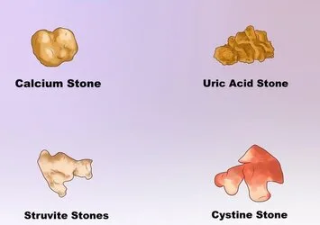
Calculus type and description
80% calcium phosphate and calcium oxalate.
Can be dotted, mulberry, or spiculated varieties
10%–20% of magnesium ammonium phosphate (struvite).
connected to microorganisms splitting urea, such as Proteus, Pseudomonas, and Klebsiella. may cause kidney stones known as staghorn stones.
5% are urate,occurs in urine that is acidic and is linked to cell lysis, small bowel illness, and gout.
Cystine
Rare (<1%). forms in the uncommon autosomal recessive metabolic disease cystinuria
Etiology
Hypercalciuria, hyperuricaemia, hypercystinuria, and hyperoxaluria are metabolic conditions.
Infection: Bacteria splitting urea, causing hyperuricemia.
Urinary stasis is caused by anatomic factors such as the horseshoe kidney and caliceal diverticulae.
Renal tubular acidosis type I, medullary sponge kidney, and adult polycystic kidney disease are examples of kidney diseases.
Medicines: indinavir, for instance.
Epidemiology
Prevalence: 2-3%, common. Risk over lifetime is 5–15%. 15% of cases are bilateral. A 3:1 male to female ratio.
History
be absent of symptoms. Severe discomfort extending from the loins to the groins caused by ureteric stones may be accompanied by nausea and vomiting.
urgency, regularity, infections, or retention of the urine.
Microscopic or frank haematuria (may not be present in up to 10% of cases).
Examination
Tenderness in the lower abdomen or loins. The most crucial differential diagnosis for elderly people to take into account is a leaking AAA. indications of systemic sepsis in the event of infection and blockage.
Investigational studies
Blood: FBC, PO4 3-, urate, Ca2+, and U&E (to evaluate renal function).
Urine: 24-hour urine collection, dipstick, microscopy, culture, and sensitivity.
CT scan (KUB film): Ninety percent of stones appear on a plain radiograph because they are radio-opaque.
Intravenous urogram: At first, a delayed dense nephrogram is seen with an obstructed ureteric stone; later images reveal a dilated pelvicaliceal system and a standing column of contrast down to the stone's location.
Non-contrast CT: Higher radiation dose, most sensitive for detecting stones.
Ultrasound: Not sensitive for tiny stones, but can demonstrate hydronephrosis or ureteral dilatation due to obstructive uropathy. used in patients who shouldn't be exposed to radiation or contrast, such as those who are pregnant.
Isotope renography: Evaluation of renal function in complex stone disease (e.g., using DTPA or DMSA).
Management
Analgesia and IV or oral hydration are the treatments for acute presentation. collecting urine to get the calculations that passed analysis. Ideal for stones that don't obstruct; most stones that are less than 5 mm can pass. Stones can be passed by ureteric relaxation with the aid of alpha-blockers, such as alfusozin. A kidney that has become infected and obstructed needs to be treated immediately. This can be done with a ureteroscopy and stent or a radiologically guided percutaneous nephrostomy along with supportive care and antibiotics.
Calculus removal: Suggested in cases of pyrexia, persistent discomfort, or blocking stones.
Ureteroscopy: A rigid or flexible ureteroscope is inserted into the bladder and up the ureter to visualize the stone, which can then be removed with a grasper, basket, or laser or ultrasonography. Urine drainage should be ensured by the placement of a JJ stent in the event that the stone is affected and cannot be removed.
Outside-the-body shock-wave lithotripsy: Non-intrusive. Calculus is subjected to an electromagnetic or piezoelectric shock wave in order to split it up into smaller pieces that can naturally pass. Fit for stones smaller than 2 cm, provided that there is no drainage blockage.
Nephrolithotomy via percutaneous means: Used for large, complicated stones, such as staghorn calculi. A nephroscope is inserted after the nephrostomy path is created, enabling the dissolution and removal of stones. During the first two days following surgery, a nephrostomy tube is left in place, and a nephrostogram is done to make sure the stone is gone and to verify ureteric drainage.
Seldom done open nephro-, pyelo-, or ureterolithotomies (for complicated cases).
Nephrectomy: in case of a non-functioning kidney, may be necessary.
Management of the cause: Based on the underlying reason, such as parathyroidectomy, dietary calcium or oxalate limitation, or allopurinol. Urate and cystine stones can be broken up by urine alkalinization with oral potassium citrate.
Suggestion: Promote a substantial intake of oral fluids.
Complications
Of stones: Septicemia, urinary retention, and infections, including pyelonephritis.
False route, perforation in ureteroscopy.
Pain, haematuria, and steinstrasse—a ureteric blockage brought on by a column of broken stone—are symptoms of lithotripsy.
Prognosis
Excellent, yet there is a 50% chance of recurrence during a five-year period, and infection may cause irreparable kidney scarring.
Definition
stone buildup in the urinary system. also referred to as renallithiasis.
Etiology
Urine becomes supersaturated with chemicals that form stones, causing crystallization around a focal point and creating calculi.
Calculus type and description
80% calcium phosphate and calcium oxalate.
Can be dotted, mulberry, or spiculated varieties
10%–20% of magnesium ammonium phosphate (struvite).
connected to microorganisms splitting urea, such as Proteus, Pseudomonas, and Klebsiella. may cause kidney stones known as staghorn stones.
5% are urate,occurs in urine that is acidic and is linked to cell lysis, small bowel illness, and gout.
Cystine
Rare (<1%). forms in the uncommon autosomal recessive metabolic disease cystinuria
Etiology
Hypercalciuria, hyperuricaemia, hypercystinuria, and hyperoxaluria are metabolic conditions.
Infection: Bacteria splitting urea, causing hyperuricemia.
Urinary stasis is caused by anatomic factors such as the horseshoe kidney and caliceal diverticulae.
Renal tubular acidosis type I, medullary sponge kidney, and adult polycystic kidney disease are examples of kidney diseases.
Medicines: indinavir, for instance.
Epidemiology
Prevalence: 2-3%, common. Risk over lifetime is 5–15%. 15% of cases are bilateral. A 3:1 male to female ratio.
History
be absent of symptoms. Severe discomfort extending from the loins to the groins caused by ureteric stones may be accompanied by nausea and vomiting.
urgency, regularity, infections, or retention of the urine.
Microscopic or frank haematuria (may not be present in up to 10% of cases).
Examination
Tenderness in the lower abdomen or loins. The most crucial differential diagnosis for elderly people to take into account is a leaking AAA. indications of systemic sepsis in the event of infection and blockage.
Investigational studies
Blood: FBC, PO4 3-, urate, Ca2+, and U&E (to evaluate renal function).
Urine: 24-hour urine collection, dipstick, microscopy, culture, and sensitivity.
CT scan (KUB film): Ninety percent of stones appear on a plain radiograph because they are radio-opaque.
Intravenous urogram: At first, a delayed dense nephrogram is seen with an obstructed ureteric stone; later images reveal a dilated pelvicaliceal system and a standing column of contrast down to the stone's location.
Non-contrast CT: Higher radiation dose, most sensitive for detecting stones.
Ultrasound: Not sensitive for tiny stones, but can demonstrate hydronephrosis or ureteral dilatation due to obstructive uropathy. used in patients who shouldn't be exposed to radiation or contrast, such as those who are pregnant.
Isotope renography: Evaluation of renal function in complex stone disease (e.g., using DTPA or DMSA).
Management
Analgesia and IV or oral hydration are the treatments for acute presentation. collecting urine to get the calculations that passed analysis. Ideal for stones that don't obstruct; most stones that are less than 5 mm can pass. Stones can be passed by ureteric relaxation with the aid of alpha-blockers, such as alfusozin. A kidney that has become infected and obstructed needs to be treated immediately. This can be done with a ureteroscopy and stent or a radiologically guided percutaneous nephrostomy along with supportive care and antibiotics.
Calculus removal: Suggested in cases of pyrexia, persistent discomfort, or blocking stones.
Ureteroscopy: A rigid or flexible ureteroscope is inserted into the bladder and up the ureter to visualize the stone, which can then be removed with a grasper, basket, or laser or ultrasonography. Urine drainage should be ensured by the placement of a JJ stent in the event that the stone is affected and cannot be removed.
Outside-the-body shock-wave lithotripsy: Non-intrusive. Calculus is subjected to an electromagnetic or piezoelectric shock wave in order to split it up into smaller pieces that can naturally pass. Fit for stones smaller than 2 cm, provided that there is no drainage blockage.
Nephrolithotomy via percutaneous means: Used for large, complicated stones, such as staghorn calculi. A nephroscope is inserted after the nephrostomy path is created, enabling the dissolution and removal of stones. During the first two days following surgery, a nephrostomy tube is left in place, and a nephrostogram is done to make sure the stone is gone and to verify ureteric drainage.
Seldom done open nephro-, pyelo-, or ureterolithotomies (for complicated cases).
Nephrectomy: in case of a non-functioning kidney, may be necessary.
Management of the cause: Based on the underlying reason, such as parathyroidectomy, dietary calcium or oxalate limitation, or allopurinol. Urate and cystine stones can be broken up by urine alkalinization with oral potassium citrate.
Suggestion: Promote a substantial intake of oral fluids.
Complications
Of stones: Septicemia, urinary retention, and infections, including pyelonephritis.
False route, perforation in ureteroscopy.
Pain, haematuria, and steinstrasse—a ureteric blockage brought on by a column of broken stone—are symptoms of lithotripsy.
Prognosis
Excellent, yet there is a 50% chance of recurrence during a five-year period, and infection may cause irreparable kidney scarring.
0 Comments
