- Published on
Surgery - Varicose Veins
Introduction
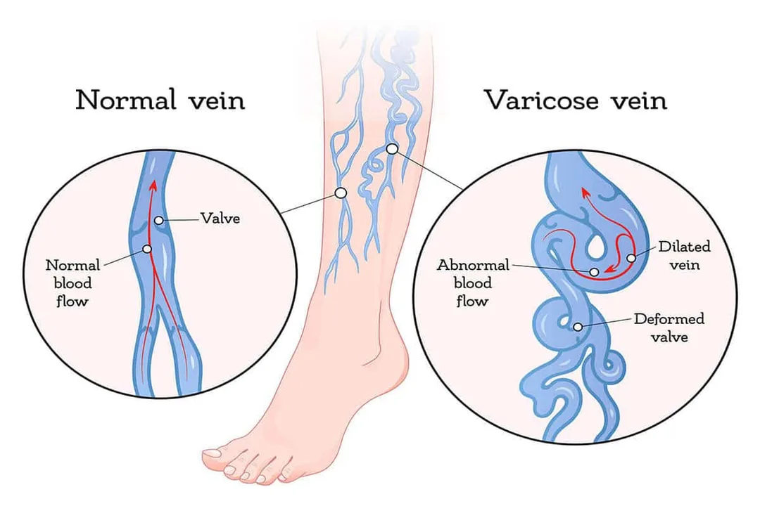
veins that have gotten longer, wider, and more twisted; these are typically the lower limbs' superficial veins. These smaller superficial venous telangiectasias and varicosities are referred to as thread veins, spider veins, or reticular veins.
Etiology
Primary: As a result of decreased elasticity, gradual dilatation, and valvular incompetence brought on by a hereditary or developmental weakening of the vein wall.
Klippel-Trenaunay syndrome (port wine stains, varicose veins, and related hypertrophic limb tissue) and Parkes Weber syndrome (like KT, but with arteriovenous fistulas) are two congenital diseases linked to varicose veins.
Secondary: Obstacles to venous outflow include ascites, lymphadenopathy, ovarian cysts, pregnancy, pelvic cancer, and retroperitoneal fibrosis.
Damage to valves: After deep vein thrombosis (DVT). Arteriovenous fistula: high flow rate.
Epidemiology
Common, rising with age; prevalence: 10-15% for men and 20-25% for women in adulthood.
History
The patient may express dissatisfaction with their outward look or report experiencing symptoms like leg pain that worsens at the end of the day or after standing for extended periods of time; swelling, itching, or consequences including ulceration, bleeding, or infection. Ask about predisposing factors, vascular risk factors, and prior history of DVT.
Examination
Examine the venous distribution, skin alterations (such as varicose eczema, lipodermatosclerosis, oedema, atrophie blanche, or ulceration) while the patient is standing.
Palpation: Incompetent perforators' sites, the facial deformities along the dilated veins, may be felt.One could feel a cough impulse across the SFJ. The tap test is a procedure whereby an impulse, which is typically absent due to competent valves, is felt distally along the vein after tapping over the SFJ. Foot pulses should be recorded if they are present. An AV fistula is indicated by the palpation of a thrill or the auscultation of a bruit.
Trendelenburg test: Capable of pinpointing valvular incompetence locations. The leg is lifted and the veins are emptied while the patient is supine. The SFJ is compressed using a hand or tourniquet. The leg is positioned dependently, and either before or after the tourniquet or hand is released, the veins are seen to be filling.
Hand Held Doppler: Capable of displaying the valvular incompetence location.
Examining the pelvis or the rectum may be necessary if secondary reasons are thought to be present.
Pathogenesis
Varicose vein etiology theories include primary valvular incompetence and the development of venous wall weakness as a result of anomalies in collagen and elastin, culminating in advanced stages of tunica media fibrosis. Hormonal fluctuations and venous hypertension are two more reasons that are implicated.
Investigations
Visualization: Duplex ultrasound: Identifies reflux or incompetence locations. Moreover, to rule out DVT (essential if surgery is being considered). MR venography: reserved for complicated situations.
Management
Conservative: Exercise recommendations (which enhance the calf muscle pump) and elevating the legs when at rest. Class II support stockings can help decrease edema and facilitate venous return.
Surgical procedures include SFJ ligation, lengthy saphenous vein stripping up to the knee, and varicosity avulsion using tiny stab incisions. To protect the sural nerve, the short saphenous vein is ligated rather than stripped.
Endovenous operations are becoming more and more common and can be carried out under local anesthesia. The long saphenous vein is ablateted using endovenous laser treatment (EVLT) after a tumescent local anesthetic is injected around the vein under ultrasound supervision.
Thermal damage and ablation are also involved in radiofrequency ablation. With foam sclerotherapy, endothelial damage, inflammation, and subsequent vein fibrosis are caused by injecting sclerosing foam along the vein with the help of ultrasound guidance. The legs are wrapped following all treatments, and early mobilization is advised.
Using laser sclerotherapy or microinjection for reticular veins and venous telangiectasia.
Complications
eczema, lipodermatosclerosis, venous ulcers, superficial thrombophlebitis, and venous pigmentation.
Treatment outcome: Recurrence. Intravenous therapy: Burns to the skin, nerve damage, bruises, emboli, and DVT. Surgical complications: bleeding, infection, nerve damage, or paraesthesia (up to 6%).
Prognosis
generally progressing slowly. After surgery, recurrence rates can reach 40%.
Introduction
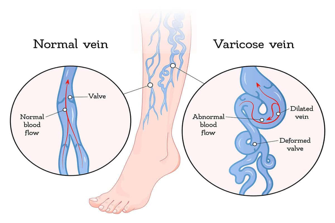
veins that have gotten longer, wider, and more twisted; these are typically the lower limbs' superficial veins. These smaller superficial venous telangiectasias and varicosities are referred to as thread veins, spider veins, or reticular veins.
Etiology
Primary: As a result of decreased elasticity, gradual dilatation, and valvular incompetence brought on by a hereditary or developmental weakening of the vein wall.
Klippel-Trenaunay syndrome (port wine stains, varicose veins, and related hypertrophic limb tissue) and Parkes Weber syndrome (like KT, but with arteriovenous fistulas) are two congenital diseases linked to varicose veins.
Secondary: Obstacles to venous outflow include ascites, lymphadenopathy, ovarian cysts, pregnancy, pelvic cancer, and retroperitoneal fibrosis.
Damage to valves: After deep vein thrombosis (DVT). Arteriovenous fistula: high flow rate.
Epidemiology
Common, rising with age; prevalence: 10-15% for men and 20-25% for women in adulthood.
History
The patient may express dissatisfaction with their outward look or report experiencing symptoms like leg pain that worsens at the end of the day or after standing for extended periods of time; swelling, itching, or consequences including ulceration, bleeding, or infection. Ask about predisposing factors, vascular risk factors, and prior history of DVT.
Examination
Examine the venous distribution, skin alterations (such as varicose eczema, lipodermatosclerosis, oedema, atrophie blanche, or ulceration) while the patient is standing.
Palpation: Incompetent perforators' sites, the facial deformities along the dilated veins, may be felt.One could feel a cough impulse across the SFJ. The tap test is a procedure whereby an impulse, which is typically absent due to competent valves, is felt distally along the vein after tapping over the SFJ. Foot pulses should be recorded if they are present. An AV fistula is indicated by the palpation of a thrill or the auscultation of a bruit.
Trendelenburg test: Capable of pinpointing valvular incompetence locations. The leg is lifted and the veins are emptied while the patient is supine. The SFJ is compressed using a hand or tourniquet. The leg is positioned dependently, and either before or after the tourniquet or hand is released, the veins are seen to be filling.
Hand Held Doppler: Capable of displaying the valvular incompetence location.
Examining the pelvis or the rectum may be necessary if secondary reasons are thought to be present.
Pathogenesis
Varicose vein etiology theories include primary valvular incompetence and the development of venous wall weakness as a result of anomalies in collagen and elastin, culminating in advanced stages of tunica media fibrosis. Hormonal fluctuations and venous hypertension are two more reasons that are implicated.
Investigations
Visualization: Duplex ultrasound: Identifies reflux or incompetence locations. Moreover, to rule out DVT (essential if surgery is being considered). MR venography: reserved for complicated situations.
Management
Conservative: Exercise recommendations (which enhance the calf muscle pump) and elevating the legs when at rest. Class II support stockings can help decrease edema and facilitate venous return.
Surgical procedures include SFJ ligation, lengthy saphenous vein stripping up to the knee, and varicosity avulsion using tiny stab incisions. To protect the sural nerve, the short saphenous vein is ligated rather than stripped.
Endovenous operations are becoming more and more common and can be carried out under local anesthesia. The long saphenous vein is ablateted using endovenous laser treatment (EVLT) after a tumescent local anesthetic is injected around the vein under ultrasound supervision.
Thermal damage and ablation are also involved in radiofrequency ablation. With foam sclerotherapy, endothelial damage, inflammation, and subsequent vein fibrosis are caused by injecting sclerosing foam along the vein with the help of ultrasound guidance. The legs are wrapped following all treatments, and early mobilization is advised.
Using laser sclerotherapy or microinjection for reticular veins and venous telangiectasia.
Complications
eczema, lipodermatosclerosis, venous ulcers, superficial thrombophlebitis, and venous pigmentation.
Treatment outcome: Recurrence. Intravenous therapy: Burns to the skin, nerve damage, bruises, emboli, and DVT. Surgical complications: bleeding, infection, nerve damage, or paraesthesia (up to 6%).
Prognosis
generally progressing slowly. After surgery, recurrence rates can reach 40%.
0 Comments
