- Published on
Dermatology - Actinic Keratosis
UVB (290–320 nm) light, in particular, causes cumulative damage to keratinocytes during prolonged and recurrent sun exposure. The onset typically occurs in middle age and is more prevalent in people who spend a lot of time outside. Actinic keratosis is uncommon in people with darker skin and common in those with lighter complexion. More often, men are impacted.
Tender lesions can form over the course of months or years. People flinch when they are ridiculed.
Damage
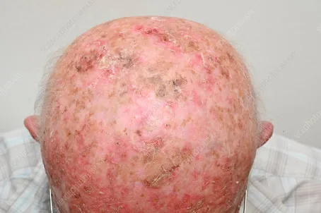
Lesions on sun-exposed areas are distinct, dry, rough, adherent, scaly macules or papules that can be solitary or many. Usually, they are accompanied with dermatoheliosis. Scale removal is a painful and challenging process. Lesions are skin-colored, brown, or yellow-brown in appearance, with a reddish undertone frequently present. The lesion is "better felt than seen" because to its rough texture. Lesions are usually circular or oval, and less than 1 cm in size.
Differential diagnosis and diagnosis
Dermatopathology confirms the clinical diagnosis. The differential comprises flat warts, squamous cell carcinoma, superficial basal cell carcinoma, seborrheic keratosis, and chronic cutaneous lupus erythematosus.
While actinic keratoses can go away on their own, they typically persist for years and have the potential to develop into squamous cell carcinoma. Applying regular UVB/UVA sunscreen will help to prevent such incidents.
Although it produces considerable erythema and erosions, topical 5-Fluorouracil (5-FU) cream 5% administered twice daily for 2-4 weeks or longer is beneficial. If administered under occlusion and/or in conjunction with topical tretinoin, efficacy is enhanced and treatment duration may be decreased.
But this causes confluent erosions, which can necessitate hospitalization. Reepithelialization happens when the course of treatment is ended. Using mild cryosurgery as a pretreatment could increase effectiveness. Although it is quite successful, imiquimod, when administered twice a week for 16 weeks, also causes irritation and erosions. Both topical retinoids and diclofenac gel, however unpleasant, are useful in treating and preventing superficial actinic keratoses and dermatioheliosis.
The use of cotton-tipped applicators or light spray cryotherapy is beneficial, particularly when paired with topical retinoids. For large lesions, facial resurfacing or peels work well. For isolated lesions, laser surgery typically works well. Photodynamic therapy is a painful and laborious yet successful treatment.
UVB (290–320 nm) light, in particular, causes cumulative damage to keratinocytes during prolonged and recurrent sun exposure. The onset typically occurs in middle age and is more prevalent in people who spend a lot of time outside. Actinic keratosis is uncommon in people with darker skin and common in those with lighter complexion. More often, men are impacted.
Tender lesions can form over the course of months or years. People flinch when they are ridiculed.
Damage
Lesions on sun-exposed areas are distinct, dry, rough, adherent, scaly macules or papules that can be solitary or many. Usually, they are accompanied with dermatoheliosis. Scale removal is a painful and challenging process. Lesions are skin-colored, brown, or yellow-brown in appearance, with a reddish undertone frequently present. The lesion is "better felt than seen" because to its rough texture. Lesions are usually circular or oval, and less than 1 cm in size.
Differential diagnosis and diagnosis
Dermatopathology confirms the clinical diagnosis. The differential comprises flat warts, squamous cell carcinoma, superficial basal cell carcinoma, seborrheic keratosis, and chronic cutaneous lupus erythematosus.
While actinic keratoses can go away on their own, they typically persist for years and have the potential to develop into squamous cell carcinoma. Applying regular UVB/UVA sunscreen will help to prevent such incidents.
Although it produces considerable erythema and erosions, topical 5-Fluorouracil (5-FU) cream 5% administered twice daily for 2-4 weeks or longer is beneficial. If administered under occlusion and/or in conjunction with topical tretinoin, efficacy is enhanced and treatment duration may be decreased.
But this causes confluent erosions, which can necessitate hospitalization. Reepithelialization happens when the course of treatment is ended. Using mild cryosurgery as a pretreatment could increase effectiveness. Although it is quite successful, imiquimod, when administered twice a week for 16 weeks, also causes irritation and erosions. Both topical retinoids and diclofenac gel, however unpleasant, are useful in treating and preventing superficial actinic keratoses and dermatioheliosis.
The use of cotton-tipped applicators or light spray cryotherapy is beneficial, particularly when paired with topical retinoids. For large lesions, facial resurfacing or peels work well. For isolated lesions, laser surgery typically works well. Photodynamic therapy is a painful and laborious yet successful treatment.
- Published on
Dermatology - Pellagra
A diet low in tryptophan, niacin, or both can cause pellagra. A diet high in maize is typically the culprit.
The three Ds of pellagra are dementia, diarrhea, and dermatitis. Pressure and solar exposure determine changes in the skin.
Damage
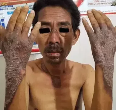
On the dorsa of the hands, neck, and face, asymmetric itching and smarting erythema is followed by breakouts of vesicles and bullae, which may burst and crust to form scaly lesions. Subsequently, the skin develops fissures, cracks, and a clear separation from normal skin. It also becomes rough, lichenified, and indurated, with dark scales and crusts covering it.
The finding of lower urine metabolite levels confirms the diagnosis.
Complete resolution is achieved by taking 100–300 mg of niacinamide orally together with other B vitamins.
A diet low in tryptophan, niacin, or both can cause pellagra. A diet high in maize is typically the culprit.
The three Ds of pellagra are dementia, diarrhea, and dermatitis. Pressure and solar exposure determine changes in the skin.
Damage
On the dorsa of the hands, neck, and face, asymmetric itching and smarting erythema is followed by breakouts of vesicles and bullae, which may burst and crust to form scaly lesions. Subsequently, the skin develops fissures, cracks, and a clear separation from normal skin. It also becomes rough, lichenified, and indurated, with dark scales and crusts covering it.
The finding of lower urine metabolite levels confirms the diagnosis.
Complete resolution is achieved by taking 100–300 mg of niacinamide orally together with other B vitamins.
- Published on
Dermatology - Tinea Capitis
Tinea Capitis
A particular class of fungus known as dermatiophytes is responsible for the infection of keratinized cutaneous tissues that causes tinea; arthrospores from these species can live for up to a year in skin scales.
The most prevalent ways for transmission are from person to person, from animals, and, less frequently, via soil. Children are the main victims of tinea capitis, a dermatophytic infection of the scalp and hair.
When tinea capitis is severe and unpleasant, it can cause painful nodules that drain pus (kerion) and cause scarring alopecia. It can also manifest as noninflammatory scaling patches, scaling and broken-off hairs. Usually, hair regrows after receiving antifungal therapy.
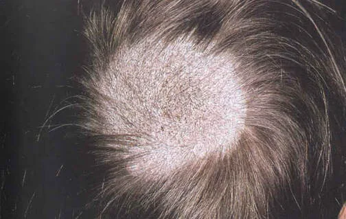
Widespread or localized alopecia is the appearance of noninflammatory scaling. The term "gray patch" refers to a partial alopecia that is typically round in shape, has fine scale and sharp borders, many broken-off hairs, and a dull gray color from an arthrospore coating. Larger patches are created when smaller ones combine. "Black dot" lesions are broken-off hairs close to the scalp that cause patients with black hair to seem to have enlarged hair shafts or "dots." The lesion is usually widespread, ill-defined, and may have low-grade folliculitis.
Kerion and favus are inflammatory masses that drain pus from numerous apertures, much like honeycomb, and are characterized by swampy, purulent, inflamed nodules and plaques that are typically unpleasant. Instead of breaking off, hairs come out and are painless to pluck. Pellicles may release pus, create sinuses, or have grains that resemble mycetomas. The surrounding hairs are matted together and have a thick crust. Although a single plaque is typical, the entire scalp may develop many lesions. Associated lymphadenopathy is often seen.
Fungal hyphae can be seen by direct microscopy of the hair shaft (collected by plucking) and scalp scales (collected with a brush covered in a drop of potassium hydroxide, or KOH). In addition to psoriasis, atopic dermatitis, lichen simplex chronicus, alopecia areata, and chronic cutaneous lupus erythematosus, the differential includes impetigo, ecthyma, crusted scabies, and kerion or favus.
Oral antidermophytes that work well are allylamines like Systemic Terbinafine 250 mg tablets.
Fluconazole 100-, 150-, or 200-mg pills, or oral suspension (10 or 40 mg/mL), are substitutes for itraconazole 100-mg capsules or oral solution (10 mg/mL).
Tinea Capitis
A particular class of fungus known as dermatiophytes is responsible for the infection of keratinized cutaneous tissues that causes tinea; arthrospores from these species can live for up to a year in skin scales.
The most prevalent ways for transmission are from person to person, from animals, and, less frequently, via soil. Children are the main victims of tinea capitis, a dermatophytic infection of the scalp and hair.
When tinea capitis is severe and unpleasant, it can cause painful nodules that drain pus (kerion) and cause scarring alopecia. It can also manifest as noninflammatory scaling patches, scaling and broken-off hairs. Usually, hair regrows after receiving antifungal therapy.
Widespread or localized alopecia is the appearance of noninflammatory scaling. The term "gray patch" refers to a partial alopecia that is typically round in shape, has fine scale and sharp borders, many broken-off hairs, and a dull gray color from an arthrospore coating. Larger patches are created when smaller ones combine. "Black dot" lesions are broken-off hairs close to the scalp that cause patients with black hair to seem to have enlarged hair shafts or "dots." The lesion is usually widespread, ill-defined, and may have low-grade folliculitis.
Kerion and favus are inflammatory masses that drain pus from numerous apertures, much like honeycomb, and are characterized by swampy, purulent, inflamed nodules and plaques that are typically unpleasant. Instead of breaking off, hairs come out and are painless to pluck. Pellicles may release pus, create sinuses, or have grains that resemble mycetomas. The surrounding hairs are matted together and have a thick crust. Although a single plaque is typical, the entire scalp may develop many lesions. Associated lymphadenopathy is often seen.
Fungal hyphae can be seen by direct microscopy of the hair shaft (collected by plucking) and scalp scales (collected with a brush covered in a drop of potassium hydroxide, or KOH). In addition to psoriasis, atopic dermatitis, lichen simplex chronicus, alopecia areata, and chronic cutaneous lupus erythematosus, the differential includes impetigo, ecthyma, crusted scabies, and kerion or favus.
Oral antidermophytes that work well are allylamines like Systemic Terbinafine 250 mg tablets.
Fluconazole 100-, 150-, or 200-mg pills, or oral suspension (10 or 40 mg/mL), are substitutes for itraconazole 100-mg capsules or oral solution (10 mg/mL).
- Published on
Dermatology - Vitiligo
Vitiligo affects 1% of people globally and can have negative social and economic repercussions for people with darker skin. Men and women experience the same chronic illness, however women seek therapy more frequently. Although it can occur at any age, the most common range for onset is 10 to 30 years. uncommonly linked to alopecia areata, pernicious anemia, and thyroid disorders.
Many patients blame illnesses, physical trauma, or psychological stress for the onset. White or prematurely gray hair are possible. In macules of vitiligo, photoaging and solar keratosis may occur.
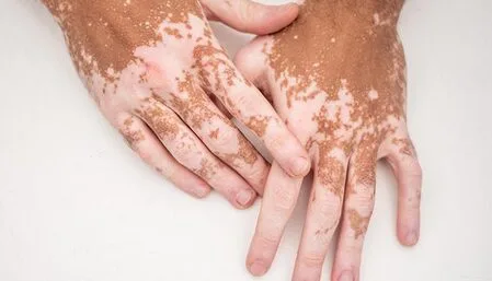
Sharply marginated, chalk-colored or pale white macules range in diameter from 5 mm to more than 5 cm, and they progressively get bigger as new ones form. Convex margins exist. The three colors of vitiligo—white, light brown, and dark brown—represent various disease phases. In a white macule, pigmentation surrounding a hair follicle indicates restored or persistent pigmentation.
Hypomelanotic macules the size of candy can also be seen. The edge of inflammatory vitiligo is more erythematous and can be itchy. A single site may have one or more macules indicative of focal vitiligo. The more prevalent type of vitiligo, known as generalized vitiligo, is typified by a broad distribution of depigmented macules that are frequently strikingly symmetrical around the mouth, eyes, fingers, elbows, and knees in addition to the low back and genital regions.
The perioral region, distal fingers and toes, lips, nipples, and genitalia are all included in the lip-tip pattern.
Large white patches are the result of confluent vitiligo, while widespread generalized vitiligo—also known as vitriligo universalis—may only leave a few regularly pigmented skin areas. Segmental vitiligo appears in a unilateral pattern, usually does not spread beyond it, and is extremely stable once it does.
Clinical diagnosis is made. Pityriasis alba, versicolor, leprosy, postinflammatory leukoderma, mycosis fungoides, chemical leukoderma, white nevi, hypomelanosis, tuberous sclerosis, Vogt-Koyanagi-Harada syndrome, Waardenburg syndrome, and piebaldism are among the conditions that fall under the differential category.
Vitiligo is unaffected by the treatment of underlying diseases. To protect the depigmented skin, use sunscreen; for people with lighter skin, this is frequently sufficient. Darker skinned people might cover up their skin tone with makeup, dyes, or self-tanning products.
Repigment local macules by applying topical psoralens, glucocorticoids, and UVA therapy.
One may try minigrafting or systemic photochemotherapy for generalized repigmentation. Alternatively, some patients choose to undergo depigmentation with bleaching chemicals, such as 20% hydroquinone monobenzylether.
Vitiligo affects 1% of people globally and can have negative social and economic repercussions for people with darker skin. Men and women experience the same chronic illness, however women seek therapy more frequently. Although it can occur at any age, the most common range for onset is 10 to 30 years. uncommonly linked to alopecia areata, pernicious anemia, and thyroid disorders.
Many patients blame illnesses, physical trauma, or psychological stress for the onset. White or prematurely gray hair are possible. In macules of vitiligo, photoaging and solar keratosis may occur.
Sharply marginated, chalk-colored or pale white macules range in diameter from 5 mm to more than 5 cm, and they progressively get bigger as new ones form. Convex margins exist. The three colors of vitiligo—white, light brown, and dark brown—represent various disease phases. In a white macule, pigmentation surrounding a hair follicle indicates restored or persistent pigmentation.
Hypomelanotic macules the size of candy can also be seen. The edge of inflammatory vitiligo is more erythematous and can be itchy. A single site may have one or more macules indicative of focal vitiligo. The more prevalent type of vitiligo, known as generalized vitiligo, is typified by a broad distribution of depigmented macules that are frequently strikingly symmetrical around the mouth, eyes, fingers, elbows, and knees in addition to the low back and genital regions.
The perioral region, distal fingers and toes, lips, nipples, and genitalia are all included in the lip-tip pattern.
Large white patches are the result of confluent vitiligo, while widespread generalized vitiligo—also known as vitriligo universalis—may only leave a few regularly pigmented skin areas. Segmental vitiligo appears in a unilateral pattern, usually does not spread beyond it, and is extremely stable once it does.
Clinical diagnosis is made. Pityriasis alba, versicolor, leprosy, postinflammatory leukoderma, mycosis fungoides, chemical leukoderma, white nevi, hypomelanosis, tuberous sclerosis, Vogt-Koyanagi-Harada syndrome, Waardenburg syndrome, and piebaldism are among the conditions that fall under the differential category.
Vitiligo is unaffected by the treatment of underlying diseases. To protect the depigmented skin, use sunscreen; for people with lighter skin, this is frequently sufficient. Darker skinned people might cover up their skin tone with makeup, dyes, or self-tanning products.
Repigment local macules by applying topical psoralens, glucocorticoids, and UVA therapy.
One may try minigrafting or systemic photochemotherapy for generalized repigmentation. Alternatively, some patients choose to undergo depigmentation with bleaching chemicals, such as 20% hydroquinone monobenzylether.
- Published on
Dermatology - Genital Penile Candidiasis
Candida species that are normally present in the normal skin flora tend to overgrow when they cause candidiasis on the nonkeratinized vaginal mucosa, or preputial sac of the penis.
There are sensitive and itchy lesions on the preputial sac and glans.
Damage
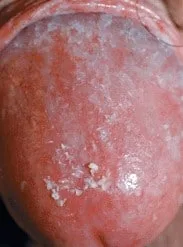
Diffuse erythema and maculopapular lesions are seen. Under the foreskin, there is edema, ulcerations, prepuce fissuring, and white plaques.
Clinical confirmation of the diagnosis is obtained using potassium hydroxide (KOH) microscopy and/or culture; lichen planus, eczema, and psoriasis are included in the differential.
Topical azole cream is the treatment; in the event of recurrences, screen and treat sexual partners as necessary.
Candida species that are normally present in the normal skin flora tend to overgrow when they cause candidiasis on the nonkeratinized vaginal mucosa, or preputial sac of the penis.
There are sensitive and itchy lesions on the preputial sac and glans.
Damage
Diffuse erythema and maculopapular lesions are seen. Under the foreskin, there is edema, ulcerations, prepuce fissuring, and white plaques.
Clinical confirmation of the diagnosis is obtained using potassium hydroxide (KOH) microscopy and/or culture; lichen planus, eczema, and psoriasis are included in the differential.
Topical azole cream is the treatment; in the event of recurrences, screen and treat sexual partners as necessary.
- Published on
Dermatology - Spitz Nevus
Spitz Nevus
Spitz nevi are somewhat common acquired lesions that typically appear in children. Because of their quick growth, they should be taken seriously.
A history of recent, quick growth (few months) is common.
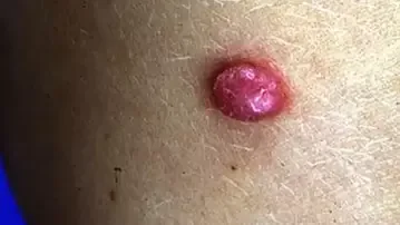
Lesions are circular, well-circumscribed, smooth-topped, hairless papules, dome-shaped nodules, or comparatively flat nodules. They are hard, typically found on the head and neck, and are typically uniformly pink-red, however they can also be tan, brown, dark brown, or even black.
Although clinical, the diagnosis needs pathological confirmation. All pink, tan, or darkly pigmented papules are included in the differential, along with moles, dysplastic nevi (amelanotic), pyogenic granuloma, hemangioma, molluscum contagiosum, juvenile xanthogranuloma, mastocytoma, and nodular melanoma.
Because the illness recurs in 10-15% of instances in lesions that have not been fully excised, thorough excision is crucial. Although spitz nevi are benign, melanoma must be checked out because of a possible histologic resemblance.
Spitz Nevus
Spitz nevi are somewhat common acquired lesions that typically appear in children. Because of their quick growth, they should be taken seriously.
A history of recent, quick growth (few months) is common.
Lesions are circular, well-circumscribed, smooth-topped, hairless papules, dome-shaped nodules, or comparatively flat nodules. They are hard, typically found on the head and neck, and are typically uniformly pink-red, however they can also be tan, brown, dark brown, or even black.
Although clinical, the diagnosis needs pathological confirmation. All pink, tan, or darkly pigmented papules are included in the differential, along with moles, dysplastic nevi (amelanotic), pyogenic granuloma, hemangioma, molluscum contagiosum, juvenile xanthogranuloma, mastocytoma, and nodular melanoma.
Because the illness recurs in 10-15% of instances in lesions that have not been fully excised, thorough excision is crucial. Although spitz nevi are benign, melanoma must be checked out because of a possible histologic resemblance.
- Published on
Dermatology - Inverse Psoriasis
Psoriasis is usually a chronic condition. In addition to environmental factors like stress, bacterial infections, trauma, or specific medications, there is a polygenic tendency. With peaks at the ages of 8, 22, and 55, onset can happen at any age. More severe disease is predicted by an early beginning. Lesions that persist for months or years affect the majority of patients.
Pruritus is prevalent, particularly in the anogenital and scalp areas
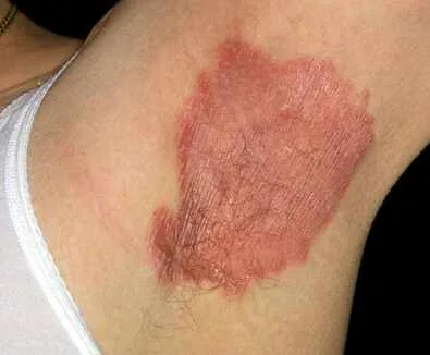
Plaques in the body folds are macerated, frequently bright red and fissured (inverse psoriasis), and lesions from intertrigo, candidiasis, or contact dermatitis can only be distinguished by a strong demarcation. In some regions, polycyclic, geographic lesions may arise from strongly marginated, dull-red plaques with loose lamellar, silvery-white scales. These could exhibit arciform, serpiginous, and annular patterns if they partially retreat. Removal of lamellar scales is simple, unless the lesion is very persistent. Lesions can be single or many, localizing to the sacralgluteal area, scalp, elbows, knees, palms, and soles—the predilection sites.
Lesions frequently spare exposed areas and exhibit bilateral symmetry. Massive hyperkeratosis with a sharply delineated foundation that is either silvery white or yellowish on the palms and soles is difficult to eliminate. Bleeding, painful fissures, and cracking are possible. Pitting, subungual hyperkeratosis, onycholysis, and yellowish-brown patches beneath the nail plate—known as the "oil spot" (pathognomonic)—are among the nail abnormalities.
Differential diagnosis and diagnosis
The differential includes seborrheic dermatitis, lichen simplex chronicus, drug eruptions, tinea, and mycosis fungoides. The diagnosis is clinical. Intertrigo, extramammary Page disease, glucagonoma syndrome, Langerhans cell histiocytosis, and Hailey-Hailey disease are among the conditions on the differential for inverse psoriasis. Onychomycosis in the nails needs to be ruled out using KOH.
Topical fluorinated glucocorticoids with occlusion can be used to treat plaques; hydrocolloid dressing works well and keeps patients from scratching. It is beneficial to inject a 3 mg/mL triamcinolone acetonide aqueous suspension diluted with normal saline into lesions that are less than 4 cm.
Although less efficacious, vitamin D analogues, tacrolimus 0.1%, and pimecrolimus 1% are good nonsteroidal agents. The use of 1% topical pimecrolimus is beneficial for inverse psoriasis.
Class II topical glucocorticoids work best when paired with tazarotene, which has comparable efficacy.
All topicals can be used in conjunction with either PUVA photochemotherapy or 311-nm UVB phototherapy.
On the scalp, tar or ketoconazole shampooing works well, but firstly, 10% salicylic acid must be used to eliminate plaques. This is followed by betamethasone valerate. Topical therapies have no effect on nail lesions.
Psoriasis is usually a chronic condition. In addition to environmental factors like stress, bacterial infections, trauma, or specific medications, there is a polygenic tendency. With peaks at the ages of 8, 22, and 55, onset can happen at any age. More severe disease is predicted by an early beginning. Lesions that persist for months or years affect the majority of patients.
Pruritus is prevalent, particularly in the anogenital and scalp areas
Plaques in the body folds are macerated, frequently bright red and fissured (inverse psoriasis), and lesions from intertrigo, candidiasis, or contact dermatitis can only be distinguished by a strong demarcation. In some regions, polycyclic, geographic lesions may arise from strongly marginated, dull-red plaques with loose lamellar, silvery-white scales. These could exhibit arciform, serpiginous, and annular patterns if they partially retreat. Removal of lamellar scales is simple, unless the lesion is very persistent. Lesions can be single or many, localizing to the sacralgluteal area, scalp, elbows, knees, palms, and soles—the predilection sites.
Lesions frequently spare exposed areas and exhibit bilateral symmetry. Massive hyperkeratosis with a sharply delineated foundation that is either silvery white or yellowish on the palms and soles is difficult to eliminate. Bleeding, painful fissures, and cracking are possible. Pitting, subungual hyperkeratosis, onycholysis, and yellowish-brown patches beneath the nail plate—known as the "oil spot" (pathognomonic)—are among the nail abnormalities.
Differential diagnosis and diagnosis
The differential includes seborrheic dermatitis, lichen simplex chronicus, drug eruptions, tinea, and mycosis fungoides. The diagnosis is clinical. Intertrigo, extramammary Page disease, glucagonoma syndrome, Langerhans cell histiocytosis, and Hailey-Hailey disease are among the conditions on the differential for inverse psoriasis. Onychomycosis in the nails needs to be ruled out using KOH.
Topical fluorinated glucocorticoids with occlusion can be used to treat plaques; hydrocolloid dressing works well and keeps patients from scratching. It is beneficial to inject a 3 mg/mL triamcinolone acetonide aqueous suspension diluted with normal saline into lesions that are less than 4 cm.
Although less efficacious, vitamin D analogues, tacrolimus 0.1%, and pimecrolimus 1% are good nonsteroidal agents. The use of 1% topical pimecrolimus is beneficial for inverse psoriasis.
Class II topical glucocorticoids work best when paired with tazarotene, which has comparable efficacy.
All topicals can be used in conjunction with either PUVA photochemotherapy or 311-nm UVB phototherapy.
On the scalp, tar or ketoconazole shampooing works well, but firstly, 10% salicylic acid must be used to eliminate plaques. This is followed by betamethasone valerate. Topical therapies have no effect on nail lesions.
- Published on
Dermatology - Atopic Dermatitis ( Eczema)
Eczema, also known as atopic dermatitis, typically first appears in infancy and peaks in early childhood with a prevalence of 15-20%. It is common for atopy, allergic rhinitis, or asthma to run in families.
Aeroallergens, including dust mites and pollen, microorganisms, autoallergens, foods—particularly eggs, milk, peanuts, soybeans, fish, and wheat—clothing, particularly wool, and bacteria are examples of triggers.
Stress may be the cause of flares, which are more frequent in the winter and after taking off clothing.
As they say, "eczema is the itch that rashes," pruritus is a must. A vicious cycle of itching, scratching, rash, itching, and scratching results from persistent scratching.
Damage
Scale/edema may be present in poorly defined erythematous patches, papules, and plaques. Scratching may cause moist crusted erosions, which, if they leak, indicate subsequent infection.
Chronic cases result in lichenification/fissures, which can be uncomfortable. People with highly pigmented skin are particularly susceptible to follicular lichenification. Infraorbital fold (Dennie-Morgan sign), periorbital pigmentation, and eyebrow alopecia can all result from rubbing.
The clinical exam and history are used to make the diagnosis. Dermatophytosis, nummular eczema, psoriasis, seborrheic dermatitis, contact dermatitis, and early mycosis fungi are examples of differential. Acrodermatitis enteropathica, glucagonoma syndrome, histidinemia, phenylketonuria, and a few immunologic illnesses are among the other uncommon conditions in the differential.
Teach patients how to apply emollients, refrain from rubbing and itching, and stop secondary infections. Apply topical hydroxyzine for pruritus, topical glucocorticoids, topical antibiotics, and wet dressings as needed. Prescribe unscented emollients, tar, hydroxyquinoline, or glucocorticoids as topical anti-inflammatories, and oil or oatmeal powder baths for subacute and chronic instances. Although they are the most effective, long-term usage of glucocorticoids may have negative effects. In cases of subacute atopic dermatitis and small flare-ups, trimecrolimus and tacrolimus are quite effective. PUVA photochemotherapy, narrow band UV (311 nm) phototherapy, and UVA-UVB phototherapy may also be successful.
Eczema, also known as atopic dermatitis, typically first appears in infancy and peaks in early childhood with a prevalence of 15-20%. It is common for atopy, allergic rhinitis, or asthma to run in families.
Aeroallergens, including dust mites and pollen, microorganisms, autoallergens, foods—particularly eggs, milk, peanuts, soybeans, fish, and wheat—clothing, particularly wool, and bacteria are examples of triggers.
Stress may be the cause of flares, which are more frequent in the winter and after taking off clothing.
As they say, "eczema is the itch that rashes," pruritus is a must. A vicious cycle of itching, scratching, rash, itching, and scratching results from persistent scratching.
Damage
Scale/edema may be present in poorly defined erythematous patches, papules, and plaques. Scratching may cause moist crusted erosions, which, if they leak, indicate subsequent infection.
Chronic cases result in lichenification/fissures, which can be uncomfortable. People with highly pigmented skin are particularly susceptible to follicular lichenification. Infraorbital fold (Dennie-Morgan sign), periorbital pigmentation, and eyebrow alopecia can all result from rubbing.
The clinical exam and history are used to make the diagnosis. Dermatophytosis, nummular eczema, psoriasis, seborrheic dermatitis, contact dermatitis, and early mycosis fungi are examples of differential. Acrodermatitis enteropathica, glucagonoma syndrome, histidinemia, phenylketonuria, and a few immunologic illnesses are among the other uncommon conditions in the differential.
Teach patients how to apply emollients, refrain from rubbing and itching, and stop secondary infections. Apply topical hydroxyzine for pruritus, topical glucocorticoids, topical antibiotics, and wet dressings as needed. Prescribe unscented emollients, tar, hydroxyquinoline, or glucocorticoids as topical anti-inflammatories, and oil or oatmeal powder baths for subacute and chronic instances. Although they are the most effective, long-term usage of glucocorticoids may have negative effects. In cases of subacute atopic dermatitis and small flare-ups, trimecrolimus and tacrolimus are quite effective. PUVA photochemotherapy, narrow band UV (311 nm) phototherapy, and UVA-UVB phototherapy may also be successful.
- Published on
Dermatology - Drug Induced Photosensitivity
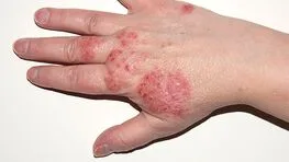
When a photoallergen and sunlight are exposed to sensitized persons, it causes a pruritic eczematous eruption that is limited to the exposed locations. This condition is clinically similar to allergic contact dermatitis. The eliciting chemical or medicine has typically been delivered topically to the patient, however systemic elicitation can also happen.
The sores are quite itchy, and they can be mistaken for allergic contact dermatitis clinically.
The lesions are crusty, scaling, vesicular, and papular. Similar to lichen planus, lichenoid eruptions can also occur occasionally. Scaling, lichenification, and severe pruritus resembling atopic dermatitis or, once more, chronic allergic contact dermatitis are symptoms of chronic drug photoallergy. The distribution pattern of photosensitivity indicates that lesions are mainly limited to light-exposed areas, while they may also spread to nearby nonexposed skin. The fact that the upper eyelids, the space beneath the nose, and the small strip of skin between the lower lip and the chin are frequently spared (shaded areas) is helpful in the diagnosis process.
When photoallergic dermatitis becomes chronic, the ailment worsens with each subsequent UV exposure and continues even when the photoallergen that caused it is removed. The consequence is extremely irritating, lichenified confluent plaques that resemble chronic dermatitis. These plaques can cause deformity and discomfort for the patient. Avoidance of photoallergens does not heal the sickness since the condition is now independent of the original photoallergen and is made worse by every new solar exposure.
The photopatch test is used to confirm the diagnosis, which is based on the patient's medical history, the eruption's pattern of allergic contact dermatitis, and the eruption's restriction to sun-exposed areas.
Sunlight and allergy avoidance are therapeutic until the illness progresses to a chronic state. Immunosuppression (azathioprine + glucocorticoids or oral cyclosporine) is necessary in severe cases.
When a photoallergen and sunlight are exposed to sensitized persons, it causes a pruritic eczematous eruption that is limited to the exposed locations. This condition is clinically similar to allergic contact dermatitis. The eliciting chemical or medicine has typically been delivered topically to the patient, however systemic elicitation can also happen.
The sores are quite itchy, and they can be mistaken for allergic contact dermatitis clinically.
The lesions are crusty, scaling, vesicular, and papular. Similar to lichen planus, lichenoid eruptions can also occur occasionally. Scaling, lichenification, and severe pruritus resembling atopic dermatitis or, once more, chronic allergic contact dermatitis are symptoms of chronic drug photoallergy. The distribution pattern of photosensitivity indicates that lesions are mainly limited to light-exposed areas, while they may also spread to nearby nonexposed skin. The fact that the upper eyelids, the space beneath the nose, and the small strip of skin between the lower lip and the chin are frequently spared (shaded areas) is helpful in the diagnosis process.
When photoallergic dermatitis becomes chronic, the ailment worsens with each subsequent UV exposure and continues even when the photoallergen that caused it is removed. The consequence is extremely irritating, lichenified confluent plaques that resemble chronic dermatitis. These plaques can cause deformity and discomfort for the patient. Avoidance of photoallergens does not heal the sickness since the condition is now independent of the original photoallergen and is made worse by every new solar exposure.
The photopatch test is used to confirm the diagnosis, which is based on the patient's medical history, the eruption's pattern of allergic contact dermatitis, and the eruption's restriction to sun-exposed areas.
Sunlight and allergy avoidance are therapeutic until the illness progresses to a chronic state. Immunosuppression (azathioprine + glucocorticoids or oral cyclosporine) is necessary in severe cases.
- Published on
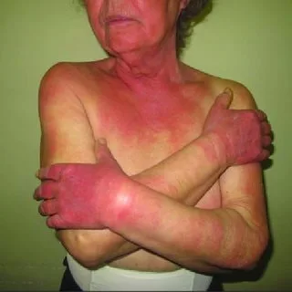
Dermatology - Mycosis Fungoides
The most prevalent cutaneous lymphoma, mycosis fungoides, usually appears in mid-to-late adulthood and affects twice as many men as women.
The illness is frequently misdiagnosed as psoriasis, nummular dermatitis, and "large plaque" parapsoriasis and can last for months to years. It's possible that pruritus exists or that no symptoms exist. Lymphadenopathy can happen, frequently following the appearance of thick plaques and nodules.
Patients may experience multiple types of lesions at the same time. Patches can be well-formed or poorly defined, randomly dispersed, and scale or nonscaling in various red hues.
These begin as superficial conditions resembling dermatophytosis, psoriasis, or eczema, and then they thicken with time. Plaques often have an oval or circular shape, however they can also be arciform, annular, or bizarrely shaped. Although lesions are spread haphazardly, they frequently spare exposed parts in the early stages. Later lesions that include nodules and tumors, either with or without ulceration, are called tumors. Leonine facies may result from extensive infiltration. Erythroderma may result from convergence. Both hair loss and palmoplantar keratoderma are present. Poikiloderma may appear right away or develop gradually.
It is challenging to diagnose in the early stages. Even though repeated samples are performed, histologic confirmation of the typical clinical lesions may not be achievable for years. There seems to be a strong correlation between lymphadenopathy and aberrant T cell circulation in the blood and internal organ involvement. The differential comprises poikiloderma, eczema, and psoriasis.
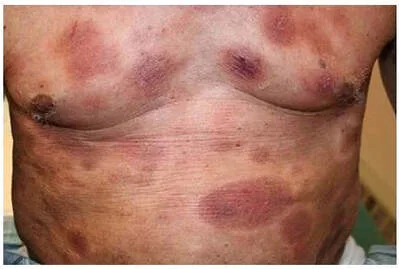
Therapy is stage-adapted and focused on symptoms. Both PUVA photochemotherapy and narrowband UVB treatment are useful when the histologic diagnosis is only compatible but not confirmed, or when there is a histologically proven plaque-stage disease without lymphadenopathy or circulating T cells. They can also be used in conjunction with subcutaneous interferon-α, oral isotretinoin, or bexarotene. Topical carmustine, topical chemotherapy with nitrogen mustard in an ointment basis (10 mg/dL), and total body electron beam therapy, either alone or in combination, are also utilized at this point. Utilize local x-ray or electron beam treatment to treat localized malignancies. The best possible combination for patients with many tumors and an extensive plaque stage is chemotherapy with electron beam plus lymphadenopathy or aberrant circulating T lymphocytes.
The most prevalent cutaneous lymphoma, mycosis fungoides, usually appears in mid-to-late adulthood and affects twice as many men as women.
The illness is frequently misdiagnosed as psoriasis, nummular dermatitis, and "large plaque" parapsoriasis and can last for months to years. It's possible that pruritus exists or that no symptoms exist. Lymphadenopathy can happen, frequently following the appearance of thick plaques and nodules.
Patients may experience multiple types of lesions at the same time. Patches can be well-formed or poorly defined, randomly dispersed, and scale or nonscaling in various red hues.
These begin as superficial conditions resembling dermatophytosis, psoriasis, or eczema, and then they thicken with time. Plaques often have an oval or circular shape, however they can also be arciform, annular, or bizarrely shaped. Although lesions are spread haphazardly, they frequently spare exposed parts in the early stages. Later lesions that include nodules and tumors, either with or without ulceration, are called tumors. Leonine facies may result from extensive infiltration. Erythroderma may result from convergence. Both hair loss and palmoplantar keratoderma are present. Poikiloderma may appear right away or develop gradually.
It is challenging to diagnose in the early stages. Even though repeated samples are performed, histologic confirmation of the typical clinical lesions may not be achievable for years. There seems to be a strong correlation between lymphadenopathy and aberrant T cell circulation in the blood and internal organ involvement. The differential comprises poikiloderma, eczema, and psoriasis.
Therapy is stage-adapted and focused on symptoms. Both PUVA photochemotherapy and narrowband UVB treatment are useful when the histologic diagnosis is only compatible but not confirmed, or when there is a histologically proven plaque-stage disease without lymphadenopathy or circulating T cells. They can also be used in conjunction with subcutaneous interferon-α, oral isotretinoin, or bexarotene. Topical carmustine, topical chemotherapy with nitrogen mustard in an ointment basis (10 mg/dL), and total body electron beam therapy, either alone or in combination, are also utilized at this point. Utilize local x-ray or electron beam treatment to treat localized malignancies. The best possible combination for patients with many tumors and an extensive plaque stage is chemotherapy with electron beam plus lymphadenopathy or aberrant circulating T lymphocytes.