- Published on
Dermatology - Non Genital Human Papillomavirus ( HPV) Infection
Human HPV is widely distributed and can cause subclinical infections, a variety of benign clinical lesions on the skin and mucous membranes (such as warts), and, more dangerously, premalignancies of the cutaneous and mucosal mucosa and squamous cell carcinoma. Keratinized skin is frequently infected by some kinds.
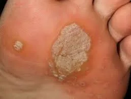
Cutaneous warts, which can range in size from tiny papules to enormous plaques (1–10 mm or greater), are distinct, benign epithelial hyperplasia with variable degrees of surface hyperkeratosis. Lesions appear at locations of minor skin injuries and are distinct, dispersed, and solitary. The pathognomonic "red or brown dots" during dermoscopy indicate thrombosed dermal papilla capillary loops. The plantar surface of the foot has a tiny, shiny papule that develops into a plaque with a rough, hyperkeratotic surface and brown or black spots scattered throughout. Tenderness may be noticeable, particularly in some acute kinds and in lesions over pressure points like the metatarsal head.
Molluscum contagiosum, seborrheic keratosis, actinic keratosis, squamous cell carcinoma in situ, syringoma, tinea versicolor, and superficial basal cell carcinoma are among the differential diagnoses that accompany the clinical diagnosis.
Given that spontaneous resolution typically takes place over the course of several months or years, aggressive interventions should generally be avoided as they can be extremely painful and leave scars. More intensive therapy are necessary for plantar warts that cause pain due to their position. There are inexpensive, very effective over-the-counter medications that cause very little discomfort. Use a 10%–20% salicylic acid and lactic acid in collodion with occlusion for small lesions, and 40% salicylic acid plaster for one week before applying the mixture in collodion for larger lesions. Options for treating resistant warts include electrosurgery, which is more successful but has a higher risk of scarring, or light cryosurgery with a cotton-tipped applicator or cryospray, which involves freezing the wart and 1-2 mm of surrounding normal tissue for about 30 seconds and repeating the procedure every four weeks.
Human HPV is widely distributed and can cause subclinical infections, a variety of benign clinical lesions on the skin and mucous membranes (such as warts), and, more dangerously, premalignancies of the cutaneous and mucosal mucosa and squamous cell carcinoma. Keratinized skin is frequently infected by some kinds.
Cutaneous warts, which can range in size from tiny papules to enormous plaques (1–10 mm or greater), are distinct, benign epithelial hyperplasia with variable degrees of surface hyperkeratosis. Lesions appear at locations of minor skin injuries and are distinct, dispersed, and solitary. The pathognomonic "red or brown dots" during dermoscopy indicate thrombosed dermal papilla capillary loops. The plantar surface of the foot has a tiny, shiny papule that develops into a plaque with a rough, hyperkeratotic surface and brown or black spots scattered throughout. Tenderness may be noticeable, particularly in some acute kinds and in lesions over pressure points like the metatarsal head.
Molluscum contagiosum, seborrheic keratosis, actinic keratosis, squamous cell carcinoma in situ, syringoma, tinea versicolor, and superficial basal cell carcinoma are among the differential diagnoses that accompany the clinical diagnosis.
Given that spontaneous resolution typically takes place over the course of several months or years, aggressive interventions should generally be avoided as they can be extremely painful and leave scars. More intensive therapy are necessary for plantar warts that cause pain due to their position. There are inexpensive, very effective over-the-counter medications that cause very little discomfort. Use a 10%–20% salicylic acid and lactic acid in collodion with occlusion for small lesions, and 40% salicylic acid plaster for one week before applying the mixture in collodion for larger lesions. Options for treating resistant warts include electrosurgery, which is more successful but has a higher risk of scarring, or light cryosurgery with a cotton-tipped applicator or cryospray, which involves freezing the wart and 1-2 mm of surrounding normal tissue for about 30 seconds and repeating the procedure every four weeks.
- Published on
Dermatology - Erythema Nodosum
Painful nodules can occur on the lower legs as a result of erythema nodosum, an essential and prevalent pattern of acute inflammation and immunologic response of the subcutaneous fat. It is the most prevalent form of panniculitis and typically affects people in their twenties and thirties, however it can strike anyone at any time. Three to six times as many women as males experience this. Many inflammatory and granulomatous disorders, including sarcoidosis, infections, and some medications can cause this.
Arthralgia(50%), most commonly in the ankle joints, fever, malaise, and painful, sensitive lesions (often lasting a few days) accompany the condition. Depending on the cause, other symptoms may manifest.
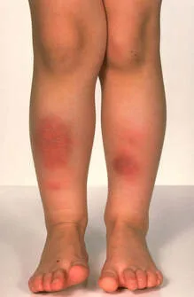
Most commonly found on the front lower legs, these indurrated, extremely sensitive nodules range in size from 3 to 20 centimeters, do not have sharp margins, and are firmly embedded in the subcutaneous fat. They are not symmetrical and tend to be bilateral. Nodules are only palpable when they are bright red to deep crimson in color.
The skin lesions are most accurately described as erythema nodosum because they have an erythematous appearance and a nodule-like texture. With time, lesions can change shape from oval to circular or arciform, and their color from green to brown to violaceous, resembling resolving hematomas. Lesions on the knees and arms are also possible, while they manifest on the face and neck quite infrequently.
Clinical criteria provide the foundation of the diagnosis, with histology providing further support as necessary.
Panarteritis nodosa, nodular vasculitis, lymphoma, nonulcerated gumma, pretibial myxedema, and any other type of panniculitis are all on the list of possible diagnoses.
New lesions emerge throughout the 6-week period of spontaneous remission.
No matter how many times a lesion heals, it will always leave a scar. Along with addressing the underlying ailment, treatment focuses on alleviating symptoms and providing support. In addition to anti-inflammatory medications, bed rest, compressive bandages (for the lower legs), and moist dressings can aid. It is only when the cause is established and infectious agents have been ruled out that systemic glucocorticoids are prescribed, despite their fast onset of action.
Painful nodules can occur on the lower legs as a result of erythema nodosum, an essential and prevalent pattern of acute inflammation and immunologic response of the subcutaneous fat. It is the most prevalent form of panniculitis and typically affects people in their twenties and thirties, however it can strike anyone at any time. Three to six times as many women as males experience this. Many inflammatory and granulomatous disorders, including sarcoidosis, infections, and some medications can cause this.
Arthralgia(50%), most commonly in the ankle joints, fever, malaise, and painful, sensitive lesions (often lasting a few days) accompany the condition. Depending on the cause, other symptoms may manifest.
Most commonly found on the front lower legs, these indurrated, extremely sensitive nodules range in size from 3 to 20 centimeters, do not have sharp margins, and are firmly embedded in the subcutaneous fat. They are not symmetrical and tend to be bilateral. Nodules are only palpable when they are bright red to deep crimson in color.
The skin lesions are most accurately described as erythema nodosum because they have an erythematous appearance and a nodule-like texture. With time, lesions can change shape from oval to circular or arciform, and their color from green to brown to violaceous, resembling resolving hematomas. Lesions on the knees and arms are also possible, while they manifest on the face and neck quite infrequently.
Clinical criteria provide the foundation of the diagnosis, with histology providing further support as necessary.
Panarteritis nodosa, nodular vasculitis, lymphoma, nonulcerated gumma, pretibial myxedema, and any other type of panniculitis are all on the list of possible diagnoses.
New lesions emerge throughout the 6-week period of spontaneous remission.
No matter how many times a lesion heals, it will always leave a scar. Along with addressing the underlying ailment, treatment focuses on alleviating symptoms and providing support. In addition to anti-inflammatory medications, bed rest, compressive bandages (for the lower legs), and moist dressings can aid. It is only when the cause is established and infectious agents have been ruled out that systemic glucocorticoids are prescribed, despite their fast onset of action.
- Published on
Dermatology - Allergic Phytodermatitis
Those who are allergic to plants can develop allergic phytodermatitis, which manifests as a linear pattern of sudden, extremely itchy, eczematous dermatitis. On the other hand, phytophotodermatitis is a type of photosensitivity reaction that can happen to anyone who gets a photosensitizing substance from plants on their skin and then goes outside. A few examples of plants that can cause allergic reactions in humans include poison ivy, poison oak, and poison sumac. Brazilian pepper, cashew, ginkgo, Indian marker, lacquer, mango, and rengas trees are among others.
Pruritis can range from very mild to quite painful, and it is common to feel its effects long before any outward changes in the skin become noticeable. Allergens have been present in the body for some time due to previous exposure to plants or foodstuffs.
Lesions
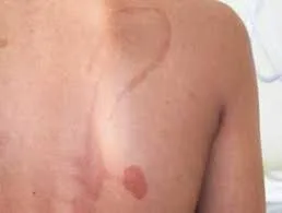
The skin becomes red in spots with clear borders; these lesions are linear in shape and can develop into papules and edematous plaques that look like cellulitis. Particularly on the face and/or genitalia, these could be quite painful. Etchings, bullae, microvesiculation, and crusts could be visible. People with darker skin tones are more likely to get postinflammatory hyperpigmentation.
History and skin examination results form the basis of a clinical diagnosis. Contact dermatitis (that isn't caused by plants), phytophotodermatitis, infections of soft tissues (cellulitis, erysipelas), atopic dermatitis, inflammatory dermatophytosis, early herpes zoster, and fixed medication eruption are all possibilities in the differential.
Wet dressings may be useful for big lesions, and topical glucocorticoids alleviate itching. If pruritis is keeping the patient from sleeping or functioning, systemic glucocorticoids may be an option to consider. Tell patients to stay away from the allergen or to immediately wash any exposed areas with water and soap if they get it.
Those who are allergic to plants can develop allergic phytodermatitis, which manifests as a linear pattern of sudden, extremely itchy, eczematous dermatitis. On the other hand, phytophotodermatitis is a type of photosensitivity reaction that can happen to anyone who gets a photosensitizing substance from plants on their skin and then goes outside. A few examples of plants that can cause allergic reactions in humans include poison ivy, poison oak, and poison sumac. Brazilian pepper, cashew, ginkgo, Indian marker, lacquer, mango, and rengas trees are among others.
Pruritis can range from very mild to quite painful, and it is common to feel its effects long before any outward changes in the skin become noticeable. Allergens have been present in the body for some time due to previous exposure to plants or foodstuffs.
Lesions
The skin becomes red in spots with clear borders; these lesions are linear in shape and can develop into papules and edematous plaques that look like cellulitis. Particularly on the face and/or genitalia, these could be quite painful. Etchings, bullae, microvesiculation, and crusts could be visible. People with darker skin tones are more likely to get postinflammatory hyperpigmentation.
History and skin examination results form the basis of a clinical diagnosis. Contact dermatitis (that isn't caused by plants), phytophotodermatitis, infections of soft tissues (cellulitis, erysipelas), atopic dermatitis, inflammatory dermatophytosis, early herpes zoster, and fixed medication eruption are all possibilities in the differential.
Wet dressings may be useful for big lesions, and topical glucocorticoids alleviate itching. If pruritis is keeping the patient from sleeping or functioning, systemic glucocorticoids may be an option to consider. Tell patients to stay away from the allergen or to immediately wash any exposed areas with water and soap if they get it.
- Published on
Dermatology - Lamellar Ichthyosis
Lamellar ichthyosis is a rare genetic skin disorder characterized by the presence of dry, scaly skin. It is inherited in an autosomal recessive manner, meaning that both parents must carry the gene for the condition in order for their child to be affected. The condition often manifests at birth and is estimated to occur in less than 1 in every 300,000 individuals.
Patients experience heat intolerance, typically during physical activity and high temperatures, as a result of the inability to perspire and excessive dehydration caused by transepidermal water loss. Children experience an elevated need for nutrients as a result of their rapid growth and peeling of the outermost layer of their skin, known as the stratum corneum.
Abnormalities
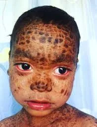
Lamellar ichthyosis manifests in newborns as a condition known as collodion baby, characterized by the infant being enveloped in a transparent membrane resembling collodion, which is shed after a few weeks. During childhood, the entire body is covered in enormous, parchment-like patches of thickened skin called hyperkeratoses. These patches can break and form a pattern that resembles tiles. The scales are extensive, thick, and brown, covering most of the body. They are particularly prominent on the lower extremities and affect the flexural areas. Verrucous hyperkeratosis can occur around the joints. Keratoderma and increased palmar/plantar creases are present on the hands and feet, and erythroderma may occur. The hair is constrained by scales and recurrent infections can lead to scarring alopecia. The presence of nail dystrophy is a result of inflammation in the nail fold. The eyes exhibit ectropion, and the lips display eclabium.
The differential diagnosis comprises X-linked ichthyosis, epidermolytic hyperkeratosis, congenital ichthyosiform erythroderma, and syndromic ichthyoses. Exclude the possibility of subsequent infection and sepsis, particularly in babies.
Infants should be admitted to the neonatal critical care unit and placed in a chamber with high humidity. They should get emolliation and be closely checked for electrolyte levels, fluid intake, and symptoms of infection. During childhood, it is important to maintain skin hydration by using emollients like hydrated petrolatum. Parents and individuals who are impacted should get instructions regarding the potential risks of overheating and heat prostration that may occur as a result of physical exercise, exposure to high environmental temperatures, and fever. Consistently applying water to the skin can partially substitute the function of sweating, resulting in the cooling of the body. As the patient matures, retinoids such as acitretin and, to a lesser extent, isotretinoin (at a dosage of 0.5–1 mg/kg) have proven to be beneficial. Nevertheless, it is imperative to consistently check blood triglyceride levels, transaminase levels, and any potential harmful effects on the bones.
Lamellar ichthyosis is a rare genetic skin disorder characterized by the presence of dry, scaly skin. It is inherited in an autosomal recessive manner, meaning that both parents must carry the gene for the condition in order for their child to be affected. The condition often manifests at birth and is estimated to occur in less than 1 in every 300,000 individuals.
Patients experience heat intolerance, typically during physical activity and high temperatures, as a result of the inability to perspire and excessive dehydration caused by transepidermal water loss. Children experience an elevated need for nutrients as a result of their rapid growth and peeling of the outermost layer of their skin, known as the stratum corneum.
Abnormalities
Lamellar ichthyosis manifests in newborns as a condition known as collodion baby, characterized by the infant being enveloped in a transparent membrane resembling collodion, which is shed after a few weeks. During childhood, the entire body is covered in enormous, parchment-like patches of thickened skin called hyperkeratoses. These patches can break and form a pattern that resembles tiles. The scales are extensive, thick, and brown, covering most of the body. They are particularly prominent on the lower extremities and affect the flexural areas. Verrucous hyperkeratosis can occur around the joints. Keratoderma and increased palmar/plantar creases are present on the hands and feet, and erythroderma may occur. The hair is constrained by scales and recurrent infections can lead to scarring alopecia. The presence of nail dystrophy is a result of inflammation in the nail fold. The eyes exhibit ectropion, and the lips display eclabium.
The differential diagnosis comprises X-linked ichthyosis, epidermolytic hyperkeratosis, congenital ichthyosiform erythroderma, and syndromic ichthyoses. Exclude the possibility of subsequent infection and sepsis, particularly in babies.
Infants should be admitted to the neonatal critical care unit and placed in a chamber with high humidity. They should get emolliation and be closely checked for electrolyte levels, fluid intake, and symptoms of infection. During childhood, it is important to maintain skin hydration by using emollients like hydrated petrolatum. Parents and individuals who are impacted should get instructions regarding the potential risks of overheating and heat prostration that may occur as a result of physical exercise, exposure to high environmental temperatures, and fever. Consistently applying water to the skin can partially substitute the function of sweating, resulting in the cooling of the body. As the patient matures, retinoids such as acitretin and, to a lesser extent, isotretinoin (at a dosage of 0.5–1 mg/kg) have proven to be beneficial. Nevertheless, it is imperative to consistently check blood triglyceride levels, transaminase levels, and any potential harmful effects on the bones.
- Published on
Dermatology - Sweet Syndrome
A rare, sudden, and recurring cutaneous reaction caused by cytokines and related with several causes. The age at which symptoms first appear is typically between 30 and 60 years, with a higher prevalence in women compared to men.
Prodromes are characterized by fever and infections in the upper respiratory tract, which may also involve gastrointestinal symptoms, tonsillitis, headache, arthralgia, and malaise. These symptoms often occur 1-3 weeks before the appearance of lesions.
Abnormalities
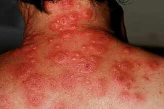
The lesions are characterized by their bright red color, smooth texture, and tenderness. They appear as little papules, about 2-4 mm in diameter, which merge together to form irregular, well-defined, inflamed plaques. Pseudovesiculation refers to a severe swelling that mimics the appearance of vesiculation. Lesions develop quickly and, as they progress, a central clearance may cause them to form round or curved patterns. Small, shallow pustules may develop. In cases where leukemia is present, the development of bullous lesions is possible and can resemble pyoderma gangrenosum.
Lesions can occur as a single or several, and they may be distributed asymmetrically or symmetrically. They are most frequently found on the face, neck, and upper extremities, but can also appear on the lower limbs. In certain cases, the lesions may be deep within the fat, resembling panniculitis or erythema nodosum. Truncal lesions are few yet prevalent, and they can manifest in a widespread manner, including generalized forms. Conjunctivitis and episcleritis may coexist.
The diagnosis is made through the assessment of clinical features, examination of tissue samples under a microscope, and confirmation with a skin biopsy. The differential diagnosis comprises erythema multiforme, erythema nodosum, prevesicular herpes simplex infection, and preulcerative pyoderma gangrenosum.
If left untreated, the lesions would gradually increase in size over a span of days or weeks and then disappear without leaving any scars. Lesions are resolved within a few days with the use of oral prednisone, administered at a dosage of 30-50 mg per day and gradually reduced over a period of 2-3 weeks. Alternatively, patients exhibit a positive response to dapsone at a dosage of 100 mg per day, or to potassium iodide. Recurrences manifest in 50% of patients, frequently affecting previously affected areas. Exclude the possibility of sepsis.
A rare, sudden, and recurring cutaneous reaction caused by cytokines and related with several causes. The age at which symptoms first appear is typically between 30 and 60 years, with a higher prevalence in women compared to men.
Prodromes are characterized by fever and infections in the upper respiratory tract, which may also involve gastrointestinal symptoms, tonsillitis, headache, arthralgia, and malaise. These symptoms often occur 1-3 weeks before the appearance of lesions.
Abnormalities
The lesions are characterized by their bright red color, smooth texture, and tenderness. They appear as little papules, about 2-4 mm in diameter, which merge together to form irregular, well-defined, inflamed plaques. Pseudovesiculation refers to a severe swelling that mimics the appearance of vesiculation. Lesions develop quickly and, as they progress, a central clearance may cause them to form round or curved patterns. Small, shallow pustules may develop. In cases where leukemia is present, the development of bullous lesions is possible and can resemble pyoderma gangrenosum.
Lesions can occur as a single or several, and they may be distributed asymmetrically or symmetrically. They are most frequently found on the face, neck, and upper extremities, but can also appear on the lower limbs. In certain cases, the lesions may be deep within the fat, resembling panniculitis or erythema nodosum. Truncal lesions are few yet prevalent, and they can manifest in a widespread manner, including generalized forms. Conjunctivitis and episcleritis may coexist.
The diagnosis is made through the assessment of clinical features, examination of tissue samples under a microscope, and confirmation with a skin biopsy. The differential diagnosis comprises erythema multiforme, erythema nodosum, prevesicular herpes simplex infection, and preulcerative pyoderma gangrenosum.
If left untreated, the lesions would gradually increase in size over a span of days or weeks and then disappear without leaving any scars. Lesions are resolved within a few days with the use of oral prednisone, administered at a dosage of 30-50 mg per day and gradually reduced over a period of 2-3 weeks. Alternatively, patients exhibit a positive response to dapsone at a dosage of 100 mg per day, or to potassium iodide. Recurrences manifest in 50% of patients, frequently affecting previously affected areas. Exclude the possibility of sepsis.
- Published on
Dermatology - Seborrheic Dermatitis
Dermatitis is a prevalent and persistent skin condition characterized by inflammation and flaking. It primarily affects areas with high sebaceous gland activity, including the face, scalp, presternal area, and body folds. Dandruff refers to mild seborrheic dermatitis occurring on the scalp. Seborrheic Dermatitis (SD) is a condition that is more prevalent in males and affects approximately 2-5% of the population. It typically begins in infancy, adolescence, or most commonly, during maturity, namely between the ages of 20 and 50.
The onset of symptoms is gradual, and in some people, symptoms worsen in cold or dry settings. Sunlight can trigger flare-ups, but it also promotes improvement in certain patients. The presence of varied pruritus is frequently exacerbated by perspiration.
Abnormalities
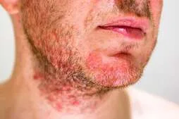
The skin can seem orange-red or gray-white, and may have greasy or white dry scaling macules, papules of different sizes (ranging from 5 to 20 mm), or patches with well-defined edges. The scalp is mostly characterized by noticeable scaling, commonly referred to as "dandruff". Lesions on the trunk are distributed in a dispersed, separate, and multi-cyclic pattern, and on the face and trunk, lesions are separate and scattered. The lesions in the body folds are widespread, exudative, well-defined, and very red in color, often accompanied by erosions and fissures.
The diagnosis is made based on clinical observations, and the possible conditions to consider include psoriasis vulgaris, impetigo, dermatophytosis, pityriasis versicolor, intertriginous candidiasis, subacute lupus erythematosus, "seborrheic" papules in secondary syphilis, Langerhans cell histiocytosis, acrodermatitis enteropathica, zinc deficiency, pemphigus, and glucagonoma syndrome.
Advise people to utilize shampoos that contain selenium sulfide, zinc pyrithione, and/or tar. Alternatively, recommend prescribing a 2% ketoconazole shampoo. The foam from these shampoos can be applied to the face and chest. In cases of high severity, apply low-potency topical glucocorticoids following shampooing. For newborns, eliminate crusts by applying warm oil compresses, then using 2% ketoconazole shampoo, and either 1–2.5% hydrocortisone cream, 2% ketoconazole cream, or 1% pimecrolimus cream. To address maintenance issues, if shampoo alone proves to be unsuccessful, utilize a solution containing 3% sulfur precipitate and 2% salicylic acid in an oil-in-water base.
Regular application of hydrocortisone cream with a concentration of 1-2.5% is effective, although it is necessary to closely observe for any signs of atrophy.
Pimecrolimus cream with a concentration of 1% and tacrolimus ointment with a concentration of 0.1% are both proven to be safe and efficacious.
Dermatitis is a prevalent and persistent skin condition characterized by inflammation and flaking. It primarily affects areas with high sebaceous gland activity, including the face, scalp, presternal area, and body folds. Dandruff refers to mild seborrheic dermatitis occurring on the scalp. Seborrheic Dermatitis (SD) is a condition that is more prevalent in males and affects approximately 2-5% of the population. It typically begins in infancy, adolescence, or most commonly, during maturity, namely between the ages of 20 and 50.
The onset of symptoms is gradual, and in some people, symptoms worsen in cold or dry settings. Sunlight can trigger flare-ups, but it also promotes improvement in certain patients. The presence of varied pruritus is frequently exacerbated by perspiration.
Abnormalities
The skin can seem orange-red or gray-white, and may have greasy or white dry scaling macules, papules of different sizes (ranging from 5 to 20 mm), or patches with well-defined edges. The scalp is mostly characterized by noticeable scaling, commonly referred to as "dandruff". Lesions on the trunk are distributed in a dispersed, separate, and multi-cyclic pattern, and on the face and trunk, lesions are separate and scattered. The lesions in the body folds are widespread, exudative, well-defined, and very red in color, often accompanied by erosions and fissures.
The diagnosis is made based on clinical observations, and the possible conditions to consider include psoriasis vulgaris, impetigo, dermatophytosis, pityriasis versicolor, intertriginous candidiasis, subacute lupus erythematosus, "seborrheic" papules in secondary syphilis, Langerhans cell histiocytosis, acrodermatitis enteropathica, zinc deficiency, pemphigus, and glucagonoma syndrome.
Advise people to utilize shampoos that contain selenium sulfide, zinc pyrithione, and/or tar. Alternatively, recommend prescribing a 2% ketoconazole shampoo. The foam from these shampoos can be applied to the face and chest. In cases of high severity, apply low-potency topical glucocorticoids following shampooing. For newborns, eliminate crusts by applying warm oil compresses, then using 2% ketoconazole shampoo, and either 1–2.5% hydrocortisone cream, 2% ketoconazole cream, or 1% pimecrolimus cream. To address maintenance issues, if shampoo alone proves to be unsuccessful, utilize a solution containing 3% sulfur precipitate and 2% salicylic acid in an oil-in-water base.
Regular application of hydrocortisone cream with a concentration of 1-2.5% is effective, although it is necessary to closely observe for any signs of atrophy.
Pimecrolimus cream with a concentration of 1% and tacrolimus ointment with a concentration of 0.1% are both proven to be safe and efficacious.
- Published on
Dermatology - Viral Exanthem
Primary systemic viral infections commonly manifest with distinctive mucocutaneous rashes, exanthems, and enanthems. An exanthem refers to a rash that appears on the skin as a result of a systemic condition. Enanthem, on the other hand, refers to mucosal lesions that are commonly connected with a systemic disorder that also causes an exanthem. These conditions are primarily caused by viral agents, although they can also result from bacterial, parasitic infections, or be triggered by drugs/toxins or autoimmune responses.
Certain viral exanthems have distinct morphologies, but, clinical symptoms alone may not always lead to an appropriate diagnosis. Obtaining a patient's medical history might provide valuable information, such as the time of year, any recent exposure to diseases, immunization records, past instances of skin rashes, and any symptoms experienced before the rash appeared. Measles is accompanied by conjunctivitis.
Prodrome refers to the early symptoms or warning signs that occur before the onset of a disease or medical condition.
The individual is experiencing an acute infection characterized by symptoms such as fever, fatigue, nasal congestion, sore throat, feelings of sickness, vomiting, diarrhea, stomach discomfort, and headache. The presence of lymphadenopathy, hepatomegaly, and splenomegaly is possible.
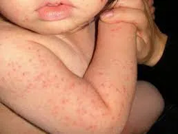
Morbilliform refers to a widespread rash that starts as individual spots and subsequently merges together. The rash typically appears in the central areas of the body, such as the head, neck, trunk, and proximal extremities, and spreads outward from there. Lesions have the potential to develop hemorrhage and exhibit petechiae. The scarlatiniform eruption manifests as a widespread and uniform redness of the skin. The vesicular eruptions first appear as small blisters with transparent fluid, which may develop into pustules. These pustules eventually detach and lead to erosions. Oropharyngeal lesions encompass several manifestations such as Koplik spots in cases of measles, petechiae on the soft palate (known as Forchheimer sign) in cytomegalovirus (CMV) or Epstein-Barr virus (EBV) infections, and microulcerative lesions in herpangina caused by coxsackie virus A.
The diagnosis is mostly based on clinical evaluation and can be further validated with serological or cultural analysis, depending on the specific requirements. Differential diagnosis encompasses cutaneous drug eruption, systemic lupus erythematosus, and Kawasaki syndrome, all of which have negative effects on the skin.
Supportive care is usually sufficient for most viral infections, however antiviral drugs might be beneficial in the early stages of the illness. Vaccination is a highly effective method for preventing most viral infections.
Primary systemic viral infections commonly manifest with distinctive mucocutaneous rashes, exanthems, and enanthems. An exanthem refers to a rash that appears on the skin as a result of a systemic condition. Enanthem, on the other hand, refers to mucosal lesions that are commonly connected with a systemic disorder that also causes an exanthem. These conditions are primarily caused by viral agents, although they can also result from bacterial, parasitic infections, or be triggered by drugs/toxins or autoimmune responses.
Certain viral exanthems have distinct morphologies, but, clinical symptoms alone may not always lead to an appropriate diagnosis. Obtaining a patient's medical history might provide valuable information, such as the time of year, any recent exposure to diseases, immunization records, past instances of skin rashes, and any symptoms experienced before the rash appeared. Measles is accompanied by conjunctivitis.
Prodrome refers to the early symptoms or warning signs that occur before the onset of a disease or medical condition.
The individual is experiencing an acute infection characterized by symptoms such as fever, fatigue, nasal congestion, sore throat, feelings of sickness, vomiting, diarrhea, stomach discomfort, and headache. The presence of lymphadenopathy, hepatomegaly, and splenomegaly is possible.
Morbilliform refers to a widespread rash that starts as individual spots and subsequently merges together. The rash typically appears in the central areas of the body, such as the head, neck, trunk, and proximal extremities, and spreads outward from there. Lesions have the potential to develop hemorrhage and exhibit petechiae. The scarlatiniform eruption manifests as a widespread and uniform redness of the skin. The vesicular eruptions first appear as small blisters with transparent fluid, which may develop into pustules. These pustules eventually detach and lead to erosions. Oropharyngeal lesions encompass several manifestations such as Koplik spots in cases of measles, petechiae on the soft palate (known as Forchheimer sign) in cytomegalovirus (CMV) or Epstein-Barr virus (EBV) infections, and microulcerative lesions in herpangina caused by coxsackie virus A.
The diagnosis is mostly based on clinical evaluation and can be further validated with serological or cultural analysis, depending on the specific requirements. Differential diagnosis encompasses cutaneous drug eruption, systemic lupus erythematosus, and Kawasaki syndrome, all of which have negative effects on the skin.
Supportive care is usually sufficient for most viral infections, however antiviral drugs might be beneficial in the early stages of the illness. Vaccination is a highly effective method for preventing most viral infections.
- Published on
Dermatology - Fissured tongue
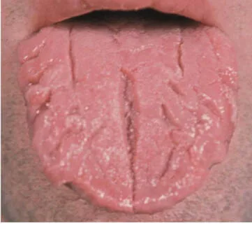
A fissured tongue is a common occurrence in approximately 11% of the population and can also be linked to psoriasis, Down syndrome, acromegaly, and Sjögren syndrome.
The absence of symptoms is observed, but the dorsal surface of the tongue has many folds arranged in an anterior-posterior direction.
The diagnosis is based only on clinical observations and assessments.
This is a typical variation and does not require any intervention.
A fissured tongue is a common occurrence in approximately 11% of the population and can also be linked to psoriasis, Down syndrome, acromegaly, and Sjögren syndrome.
The absence of symptoms is observed, but the dorsal surface of the tongue has many folds arranged in an anterior-posterior direction.
The diagnosis is based only on clinical observations and assessments.
This is a typical variation and does not require any intervention.
- Published on
Dermatology - Nevus of Ota
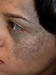
Nevus of Ota is a pigmented skin abnormality that can either be present at birth or develop in early childhood or during puberty. It is prevalent among individuals of Asian heritage, however it is not exclusive to them.
The pigmentation can vary in intensity, ranging from modest to highly disfiguring. It can affect either one side or both sides of the body, and is characterized by a patchy, dark mixture of blue and brown hyperpigmentation on the skin. The condition mostly affects the skin and mucous membranes that are supplied by the first and second divisions of the trigeminal nerve. The blue coloration is caused by the existence of ectopic melanocytes within the dermis.
The diagnosis is established through clinical means.
Laser treatment is a highly effective method for addressing this disfiguring condition.
Nevus of Ota is a pigmented skin abnormality that can either be present at birth or develop in early childhood or during puberty. It is prevalent among individuals of Asian heritage, however it is not exclusive to them.
The pigmentation can vary in intensity, ranging from modest to highly disfiguring. It can affect either one side or both sides of the body, and is characterized by a patchy, dark mixture of blue and brown hyperpigmentation on the skin. The condition mostly affects the skin and mucous membranes that are supplied by the first and second divisions of the trigeminal nerve. The blue coloration is caused by the existence of ectopic melanocytes within the dermis.
The diagnosis is established through clinical means.
Laser treatment is a highly effective method for addressing this disfiguring condition.
- Published on
Dermatology - Chancroid
Haemophilus ducreyi, a gram-negative streptobacillus that is endemic in tropical and subtropical underdeveloped countries but rare in industrialized ones, is the cause of chancroid.
Four to seven days after exposure, a primary infection (break in epithelium) occurs at the site of inoculation, followed by lymphadenitis. Prepuce edema is frequent. Fifty percent of patients experience a painful, typically unilateral, gingual lymphadenitis 7–21 days following the original lesion.
A painful and irritable papule with an erythematous halo develops into an erosion, pustule, and ulcer. The base of the ulcer is friable with granulation tissue and covered in gray to yellow exudate. The ulcer's edges are sharp, undermined, and not indurated.
One or more ulcers may combine to become massive, serpiginous ulcers that are larger than two centimeters. An ulcer could heal before a buboe appears. Buboes can drain on their own and have erythema overlaying them.
One-third of patients had tender lymphadenopathy along with a painful ulcer, which suggests chancroid. The identification of H. ducreyi on specialized culture media is necessary for a conclusive diagnosis. Eliminate co-infections with T. pallidum, herpes simplex virus, and HIV. The differential include lymphogranuloma venereum, genital herpes, syphilis, trauma, inguinal hernia, plague, and tularemia.
Options for treatment include intramuscular ceftriaxone in a single dosage, ciprofloxacin 500 mg twice daily for three days (not recommended in pregnancy), erythromycin base 500 mg three times daily for seven days, and azithromycin 1 g in a single dose. There have been reports of erythromycin and ciprofloxacin resistance.
Haemophilus ducreyi, a gram-negative streptobacillus that is endemic in tropical and subtropical underdeveloped countries but rare in industrialized ones, is the cause of chancroid.
Four to seven days after exposure, a primary infection (break in epithelium) occurs at the site of inoculation, followed by lymphadenitis. Prepuce edema is frequent. Fifty percent of patients experience a painful, typically unilateral, gingual lymphadenitis 7–21 days following the original lesion.
A painful and irritable papule with an erythematous halo develops into an erosion, pustule, and ulcer. The base of the ulcer is friable with granulation tissue and covered in gray to yellow exudate. The ulcer's edges are sharp, undermined, and not indurated.
One or more ulcers may combine to become massive, serpiginous ulcers that are larger than two centimeters. An ulcer could heal before a buboe appears. Buboes can drain on their own and have erythema overlaying them.
One-third of patients had tender lymphadenopathy along with a painful ulcer, which suggests chancroid. The identification of H. ducreyi on specialized culture media is necessary for a conclusive diagnosis. Eliminate co-infections with T. pallidum, herpes simplex virus, and HIV. The differential include lymphogranuloma venereum, genital herpes, syphilis, trauma, inguinal hernia, plague, and tularemia.
Options for treatment include intramuscular ceftriaxone in a single dosage, ciprofloxacin 500 mg twice daily for three days (not recommended in pregnancy), erythromycin base 500 mg three times daily for seven days, and azithromycin 1 g in a single dose. There have been reports of erythromycin and ciprofloxacin resistance.