- Published on
Dermatology - Oropharyngeal Lichen Planus
Lichen planus manifestations in the oropharynx
Approximately 40-60% of individuals diagnosed with lichen planus experience involvement in the oropharynx. Lichen planus is a dermatosis characterized by acute or chronic inflammation. It often occurs in individuals between the ages of 30 and 60, with a higher frequency in women. Idiopathic is the most common cause, however cell-mediated immunity can have a significant impact.
The onset can be either sudden (occurring within a few days) or gradual (taking place over several weeks), and the lesions persist for a duration of months to years. Lesions can be either asymptomatic or pruritic, and mucous membrane lesions are particularly unpleasant, especially when they are ulcerated. Postinflammatory hyperpigmentation is a frequent occurrence. Lesions may appear on the lips and in a linear pattern following trauma, known as the Koebner or isomorphic phenomena.
Abnormalities
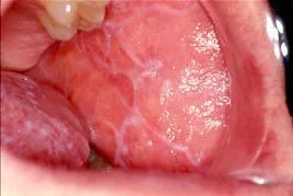
Skin lesions exhibit the following characteristics: they have a color ranging from pink to purple, they are shiny, they cause itching (pruritic), and they have a polygonal shape. These lesions are in the form of papules, which have a diameter between 1 and 10 mm. The oropharyngeal cavity has milky-white papules known as Wickham striae. These striae are characterized by a reticulate pattern of lacy-white hyperkeratosis, leukoplakia, or atrophy. The tongue and buccal mucosa may have superficial erosions covered by fibrin clots, which can cause pain. Additionally, intact blisters and bright red gingiva may be present.
The diagnosis is established through clinical examination and subsequently confirmed via biopsy. The differential diagnosis for oropharyngeal lesions encompasses leukoplakia, candidiasis, lupus, trauma, secondary syphilis, pemphigus vulgaris, and bullous pemphigoid.
Apply topical glucocorticoids with occlusion for skin lesions and use intralesional triamcinolone (3 mg/mL) for lesions in the mouth and lips. For very symptomatic oral lesions, use cyclosporine and tacrolimus solutions as a mouthwash.
Systemic
For very resistant and widespread instances, a daily oral dose of 5 mg/kg of cyclosporine can effectively bring about a quick improvement, with a minimal likelihood of the condition returning. Oral prednisone is beneficial for those experiencing symptomatic pruritus, painful erosions, dysphagia, or cosmetic deformity. An abbreviated, gradually decreasing regimen is recommended: 70 mg as an initial dose, followed by a daily reduction of 5 mg. Retinoids, specifically Acitretin, at a dosage of 1 mg/kg per day, can be beneficial as a supplementary treatment in severe instances. However, additional topical treatment is necessary. Photochemotherapy can be beneficial for those who do not respond well to both topical and systemic treatments.
Lichen planus manifestations in the oropharynx
Approximately 40-60% of individuals diagnosed with lichen planus experience involvement in the oropharynx. Lichen planus is a dermatosis characterized by acute or chronic inflammation. It often occurs in individuals between the ages of 30 and 60, with a higher frequency in women. Idiopathic is the most common cause, however cell-mediated immunity can have a significant impact.
The onset can be either sudden (occurring within a few days) or gradual (taking place over several weeks), and the lesions persist for a duration of months to years. Lesions can be either asymptomatic or pruritic, and mucous membrane lesions are particularly unpleasant, especially when they are ulcerated. Postinflammatory hyperpigmentation is a frequent occurrence. Lesions may appear on the lips and in a linear pattern following trauma, known as the Koebner or isomorphic phenomena.
Abnormalities
Skin lesions exhibit the following characteristics: they have a color ranging from pink to purple, they are shiny, they cause itching (pruritic), and they have a polygonal shape. These lesions are in the form of papules, which have a diameter between 1 and 10 mm. The oropharyngeal cavity has milky-white papules known as Wickham striae. These striae are characterized by a reticulate pattern of lacy-white hyperkeratosis, leukoplakia, or atrophy. The tongue and buccal mucosa may have superficial erosions covered by fibrin clots, which can cause pain. Additionally, intact blisters and bright red gingiva may be present.
The diagnosis is established through clinical examination and subsequently confirmed via biopsy. The differential diagnosis for oropharyngeal lesions encompasses leukoplakia, candidiasis, lupus, trauma, secondary syphilis, pemphigus vulgaris, and bullous pemphigoid.
Apply topical glucocorticoids with occlusion for skin lesions and use intralesional triamcinolone (3 mg/mL) for lesions in the mouth and lips. For very symptomatic oral lesions, use cyclosporine and tacrolimus solutions as a mouthwash.
Systemic
For very resistant and widespread instances, a daily oral dose of 5 mg/kg of cyclosporine can effectively bring about a quick improvement, with a minimal likelihood of the condition returning. Oral prednisone is beneficial for those experiencing symptomatic pruritus, painful erosions, dysphagia, or cosmetic deformity. An abbreviated, gradually decreasing regimen is recommended: 70 mg as an initial dose, followed by a daily reduction of 5 mg. Retinoids, specifically Acitretin, at a dosage of 1 mg/kg per day, can be beneficial as a supplementary treatment in severe instances. However, additional topical treatment is necessary. Photochemotherapy can be beneficial for those who do not respond well to both topical and systemic treatments.
- Published on
Dermatology - Grover Disease
Grover's disease
Grover's illness is a skin condition characterized by itching and resembling Darier's disease. However, it does not have a known genetic propensity and is typically temporary. Adult onset often occurs around the age of 50, with a higher prevalence in men compared to women. Precipitating factors encompass intense physical exertion that induces sweating, exposure to sun radiation, high temperatures, prolonged fever, and individuals confined to bed. Alternative term: transitory acantholytic dermatosis.The primary symptom is pruritus.
Abnormalities
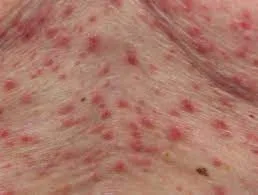
The individual has developed distinct papular or papulovesicular lesions, which might range from few to many in number. When touched, the surface might feel either smooth or warty.
The diagnosis is made based on clinical observations, and the possible conditions to consider are eczema, folliculitis, solar lentigo, solar keratosis, scabies, miliaria, Darier disease, Hailey-Hailey disease, and pemphigus vulgaris.
The recommended treatment involves the use of glucocorticosteroids applied under occlusion, as well as UVB or PUVA photochemotherapy.
Administer oral glucocorticosteroids, dapsone, and isotretinoin to patients who do not respond to other treatments.
Grover's disease
Grover's illness is a skin condition characterized by itching and resembling Darier's disease. However, it does not have a known genetic propensity and is typically temporary. Adult onset often occurs around the age of 50, with a higher prevalence in men compared to women. Precipitating factors encompass intense physical exertion that induces sweating, exposure to sun radiation, high temperatures, prolonged fever, and individuals confined to bed. Alternative term: transitory acantholytic dermatosis.The primary symptom is pruritus.
Abnormalities
The individual has developed distinct papular or papulovesicular lesions, which might range from few to many in number. When touched, the surface might feel either smooth or warty.
The diagnosis is made based on clinical observations, and the possible conditions to consider are eczema, folliculitis, solar lentigo, solar keratosis, scabies, miliaria, Darier disease, Hailey-Hailey disease, and pemphigus vulgaris.
The recommended treatment involves the use of glucocorticosteroids applied under occlusion, as well as UVB or PUVA photochemotherapy.
Administer oral glucocorticosteroids, dapsone, and isotretinoin to patients who do not respond to other treatments.
- Published on
Dermatology - Herpes Zoster
Herpes Zoster
Acute dermatomal recurrence of varicella-zoster virus (VZV), commonly referred to as shingles or herpes zoster, is typified by intense unilateral pain and a vesiculobullous reaction. Phrenic neuralgia (PHN) is a significant morbidity.
Three separate clinical stages are present in herpes zoster: prodrome, active infection, and postherpetic neuralgia. Angina or an acute abdomen can be mimicked by the pain and tenderness in the affected dermatome during the prodrome. There can be zoster without rash.
There may be flu-like symptoms, such as lymphadenopathy. Lesions appear in waves during the active infection, lasting roughly a week each, and then subside in two to four weeks. There may be temperature, pain, and touch sensory nerve abnormalities as well as (mild) motor paralysis (Bell Palsy). Complications include uveitis, keratitis, conjunctivitis, retinitis, optic neuritis, glaucoma, proptosis, cicatricial lid retraction, and extraocular muscular palsies can arise from ocular involvement. After ocular involvement, delayed contralateral hemiparesis may also develop, presenting with hemiplegia and headache.
Constant, intense, stabbing, or scorching pain that lasts for months or years is known as postherpetic neuralgia.
Damage
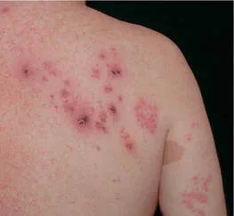
On an edematous base, the lesions are erythematous. Clear, clustered vesicles are superimposed; these can occasionally bleed and degrade to produce crusted lesions. The dermatome innervated by the sensory ganglion where the infection was reactivated is the only area where lesions can occur.
The thoracic (>50%), trigeminal (10–20%), lumbosacral, and cervical (10–20%) dermatomes are the most common locations for them. Depending on the dermatome involved, vesicles and erosions may appear in the mouth, vagina, and bladder.
The Tzanck test, DFA, or viral culture can be used to confirm the clinical diagnosis and rule out HSV infection. The prodrome can mimic vertigo, acute abdominal, cardiac or pleural illness, or migraine. Phytoallergy (poison ivy) and herpes simplex virus (HSV) infection are two differential diagnoses for the lesions.
If antiviral therapy is initiated early in the course of the illness, it is successful. Give oral valacyclovir 1 g every 8 hours, acyclovir 800 mg five times a day for seven days, or famciclovir 500 mg every eight hours. For individuals who are not significantly immunocompromised, extend treatment to 10 days and provide intravenous acylclovir or foscarnet. Gabapentin, pregabalin, tricyclic antidepressants, capaicin cream, and nerve block are useful for treating postherpetic neuralgia.
Herpes Zoster
Acute dermatomal recurrence of varicella-zoster virus (VZV), commonly referred to as shingles or herpes zoster, is typified by intense unilateral pain and a vesiculobullous reaction. Phrenic neuralgia (PHN) is a significant morbidity.
Three separate clinical stages are present in herpes zoster: prodrome, active infection, and postherpetic neuralgia. Angina or an acute abdomen can be mimicked by the pain and tenderness in the affected dermatome during the prodrome. There can be zoster without rash.
There may be flu-like symptoms, such as lymphadenopathy. Lesions appear in waves during the active infection, lasting roughly a week each, and then subside in two to four weeks. There may be temperature, pain, and touch sensory nerve abnormalities as well as (mild) motor paralysis (Bell Palsy). Complications include uveitis, keratitis, conjunctivitis, retinitis, optic neuritis, glaucoma, proptosis, cicatricial lid retraction, and extraocular muscular palsies can arise from ocular involvement. After ocular involvement, delayed contralateral hemiparesis may also develop, presenting with hemiplegia and headache.
Constant, intense, stabbing, or scorching pain that lasts for months or years is known as postherpetic neuralgia.
Damage
On an edematous base, the lesions are erythematous. Clear, clustered vesicles are superimposed; these can occasionally bleed and degrade to produce crusted lesions. The dermatome innervated by the sensory ganglion where the infection was reactivated is the only area where lesions can occur.
The thoracic (>50%), trigeminal (10–20%), lumbosacral, and cervical (10–20%) dermatomes are the most common locations for them. Depending on the dermatome involved, vesicles and erosions may appear in the mouth, vagina, and bladder.
The Tzanck test, DFA, or viral culture can be used to confirm the clinical diagnosis and rule out HSV infection. The prodrome can mimic vertigo, acute abdominal, cardiac or pleural illness, or migraine. Phytoallergy (poison ivy) and herpes simplex virus (HSV) infection are two differential diagnoses for the lesions.
If antiviral therapy is initiated early in the course of the illness, it is successful. Give oral valacyclovir 1 g every 8 hours, acyclovir 800 mg five times a day for seven days, or famciclovir 500 mg every eight hours. For individuals who are not significantly immunocompromised, extend treatment to 10 days and provide intravenous acylclovir or foscarnet. Gabapentin, pregabalin, tricyclic antidepressants, capaicin cream, and nerve block are useful for treating postherpetic neuralgia.
- Published on
Dermatology - Hemangioma of Infancy
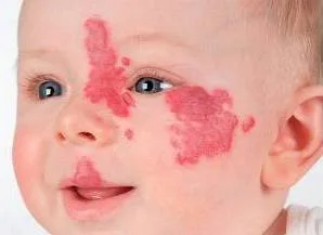
The most common tumor of infancy, known as strawberry hemangioma or infantile hemangioma, affects 1-2.5% of infants and up to 10% of white children by the time they are a year old. More girls than boys are impacted (3:1).Infantile hemangiomas often grow quickly in the first year of life and then gradually recede during the following one to five years due to an involution process. It is often finished by the time an individual reaches the age of ten. Involution varies widely and is unrelated to size, location, or appearance.
Infantile hemangiomas are 1–8 cm in diameter, soft, compressible nodules or plaques that range in color from brilliant red to deep purple. A white to gray central region emerges as involution progresses, and ulceration could happen. Lesions are typically isolated, localized, or encompass a whole area; they most frequently occur on the head and neck (50%) or the trunk (25%), while they can also occur on the extremities and oral mucosa.
Clinical and MRI results showing sluggish blood flow across the lesion serve as the basis for the diagnosis.
Vascular malformation is ruled out by GLUT-1 immunoreactivity.
Since the majority of hemangiomas spontaneously involute with minimal lingering skin alteration at the location, the best course of action for most lesions is active nonintervention, which also yields the greatest cosmetic outcome. About 25% of lesions that ulcerate or block important tissues like the throat, ears, or eyes require treatment. Systemic treatment is challenging and necessitates knowledge and skill.
Medical and surgical interventions encompass intralesional and systemic high-dose glucocorticoids, cryosurgery, interferon α (IFN-α), propanolol, and continuous wave or pulsed dye laser.
The most common tumor of infancy, known as strawberry hemangioma or infantile hemangioma, affects 1-2.5% of infants and up to 10% of white children by the time they are a year old. More girls than boys are impacted (3:1).Infantile hemangiomas often grow quickly in the first year of life and then gradually recede during the following one to five years due to an involution process. It is often finished by the time an individual reaches the age of ten. Involution varies widely and is unrelated to size, location, or appearance.
Infantile hemangiomas are 1–8 cm in diameter, soft, compressible nodules or plaques that range in color from brilliant red to deep purple. A white to gray central region emerges as involution progresses, and ulceration could happen. Lesions are typically isolated, localized, or encompass a whole area; they most frequently occur on the head and neck (50%) or the trunk (25%), while they can also occur on the extremities and oral mucosa.
Clinical and MRI results showing sluggish blood flow across the lesion serve as the basis for the diagnosis.
Vascular malformation is ruled out by GLUT-1 immunoreactivity.
Since the majority of hemangiomas spontaneously involute with minimal lingering skin alteration at the location, the best course of action for most lesions is active nonintervention, which also yields the greatest cosmetic outcome. About 25% of lesions that ulcerate or block important tissues like the throat, ears, or eyes require treatment. Systemic treatment is challenging and necessitates knowledge and skill.
Medical and surgical interventions encompass intralesional and systemic high-dose glucocorticoids, cryosurgery, interferon α (IFN-α), propanolol, and continuous wave or pulsed dye laser.
- Published on
Dermatology - Hidradenitis Suppurativa
After puberty,hidradenitis suppurativa strikes, affecting women more frequently than men (about 4% of women). Men are more likely to experience anogenital involvement than women are axillary involvement. Predispositions include smoking, obesity, and a family history of HS or acne.
varies in severity from tiny red nodules that heal on their own to significant morbidity brought on by persistent discomfort, draining sinuses, scarring, and limited movement. Ages over 35 are typically when spontaneous remission occurs, however they don't always.
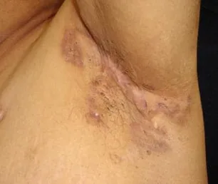
Open and double comedones evolve into extremely sensitive, red, inflamed nodules or abscesses that drain or resolve purulent or seropurulent material, leading to the development of sensitive sinus tracts. Fibrosis, "bridge" scars, hypertrophic and keloidal scars, contractures, and in rare cases, lymphedema of the related limb come next. The axillae, breasts, anogenital region, and groin are the common locations for lesions, which can also affect the scalp, buttocks, perineum involving the scrotum or vulva, and entire back. Lesions are frequently bilateral.
The differential diagnosis consists of furuncle, carbuncle, lymphadenitis, ruptured inclusion cyst, lymphogranuloma venereum, also known as cat-scratch disease, actinomycosis, donovanosis, scrofuloderma, ulcerative colitis, and regional enteritis. The diagnosis is clinical.
The treatment for this condition includes intralesional glucocorticoids, surgery, isotretinoin, and systemic antibiotics such as erythromycin (250–500 mg four times a day), tetracycline (250–500 mg four times a day), minocycline (100 mg twice a day), or clindamycin (300 mg twice a day), along with rifampin (300 mg twice a day). Triamcinolone (3–5 mg/mL) injected intralesionally, followed by incision and drainage, is used to treat painful nodules and abscesses. Remove and drain any persistent, recurrent fibrotic nodules or sinus tracts; split skin grafting may be required for the full excision of the axilla or other affected anogenital region. Use 70 mg of prednisone per day for two to three days, tapering over a period of 14 days, for severe discomfort and inflammation. Oral isotretinoin may avoid follicular plugging even if it is ineffective in cases of severe illness and should be taken in conjunction with surgical excision. Depression is widespread and has to be managed.
After puberty,hidradenitis suppurativa strikes, affecting women more frequently than men (about 4% of women). Men are more likely to experience anogenital involvement than women are axillary involvement. Predispositions include smoking, obesity, and a family history of HS or acne.
varies in severity from tiny red nodules that heal on their own to significant morbidity brought on by persistent discomfort, draining sinuses, scarring, and limited movement. Ages over 35 are typically when spontaneous remission occurs, however they don't always.
Open and double comedones evolve into extremely sensitive, red, inflamed nodules or abscesses that drain or resolve purulent or seropurulent material, leading to the development of sensitive sinus tracts. Fibrosis, "bridge" scars, hypertrophic and keloidal scars, contractures, and in rare cases, lymphedema of the related limb come next. The axillae, breasts, anogenital region, and groin are the common locations for lesions, which can also affect the scalp, buttocks, perineum involving the scrotum or vulva, and entire back. Lesions are frequently bilateral.
The differential diagnosis consists of furuncle, carbuncle, lymphadenitis, ruptured inclusion cyst, lymphogranuloma venereum, also known as cat-scratch disease, actinomycosis, donovanosis, scrofuloderma, ulcerative colitis, and regional enteritis. The diagnosis is clinical.
The treatment for this condition includes intralesional glucocorticoids, surgery, isotretinoin, and systemic antibiotics such as erythromycin (250–500 mg four times a day), tetracycline (250–500 mg four times a day), minocycline (100 mg twice a day), or clindamycin (300 mg twice a day), along with rifampin (300 mg twice a day). Triamcinolone (3–5 mg/mL) injected intralesionally, followed by incision and drainage, is used to treat painful nodules and abscesses. Remove and drain any persistent, recurrent fibrotic nodules or sinus tracts; split skin grafting may be required for the full excision of the axilla or other affected anogenital region. Use 70 mg of prednisone per day for two to three days, tapering over a period of 14 days, for severe discomfort and inflammation. Oral isotretinoin may avoid follicular plugging even if it is ineffective in cases of severe illness and should be taken in conjunction with surgical excision. Depression is widespread and has to be managed.
- Published on
Dermatology - Staphylococcal Scalded Skin Syndrome
In infants and young children, Staphylococcus aureus generates exfoliating toxins that cause the skin to peel off. After the synthesis, absorption, and subsequent host response that the poison triggered, illness develops.
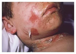
The localized form is characterized by intact, clustered flaccid purulent bullae that, when burst, produce crusty or wet red erosions. In an intertriginous area, lesions are frequently grouped together. In the generalized form, the earliest symptoms include a fine, stippled, sandpaper-like appearance, a widespread, ill-defined erythema, or a macular scarlatiniform rash (staphylococcal scarlet fever syndrome). The affected area turns painful and the erythema develops during the course of a day. The rash starts out on the face, neck, axillae, and groins before spreading over the course of 24 to 48 hours.
Subsequent sloughing of the epidermis's surface layers is especially noticeable on the face and at the pressure points located in the flexural areas of the neck, axillae, groins, and back. Epidermolysis is characterized by wrinkled epidermis that can be removed with light pressure (skin feels like wet tissue paper) (Nikolsky sign). Some babies have flaccid bullae, which become red, wet erosions when they are unroofed. Desquamation happens when the body heals.
Clinical diagnosis is made, and bacterial culture confirms the diagnosis. Scarlet fever, severe cutaneous medication eruption, and Kawasaki syndrome are among the differential diagnoses.
Administer systemic antibiotics to address infection and prevent the generation of toxins.
In infants and young children, Staphylococcus aureus generates exfoliating toxins that cause the skin to peel off. After the synthesis, absorption, and subsequent host response that the poison triggered, illness develops.
The localized form is characterized by intact, clustered flaccid purulent bullae that, when burst, produce crusty or wet red erosions. In an intertriginous area, lesions are frequently grouped together. In the generalized form, the earliest symptoms include a fine, stippled, sandpaper-like appearance, a widespread, ill-defined erythema, or a macular scarlatiniform rash (staphylococcal scarlet fever syndrome). The affected area turns painful and the erythema develops during the course of a day. The rash starts out on the face, neck, axillae, and groins before spreading over the course of 24 to 48 hours.
Subsequent sloughing of the epidermis's surface layers is especially noticeable on the face and at the pressure points located in the flexural areas of the neck, axillae, groins, and back. Epidermolysis is characterized by wrinkled epidermis that can be removed with light pressure (skin feels like wet tissue paper) (Nikolsky sign). Some babies have flaccid bullae, which become red, wet erosions when they are unroofed. Desquamation happens when the body heals.
Clinical diagnosis is made, and bacterial culture confirms the diagnosis. Scarlet fever, severe cutaneous medication eruption, and Kawasaki syndrome are among the differential diagnoses.
Administer systemic antibiotics to address infection and prevent the generation of toxins.
- Published on
Dermatology - Polymorphous Light Eruption (PMLE)
A collection of varied, idiopathic, acquired, acute recurring eruptions that are a delayed reaction to UV radiation are together referred to as PMLEs. The most prevalent type of photodermatitis, PMLE often appears in the third decade of life. It is more prevalent among lighter-skinned people and women than in men. Among American Indians, both in North and South America, actinic prurigo is a hereditary type.
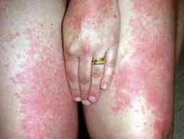
Early spring and summer are the times for lesions to appear. These include erythematous macules, papules, plaques, and vesicles. Nonetheless, there is only ever one form of eruption in each patient, most often papular or papulovesicular. After exposure, PMLE often manifests within hours and lasts for seven to ten days. Often, the eruptions stop occurring by the end of the summer, indicating a "hardening." Paresthesia (tingling) and pruritus (which may occur before the rash appears) are the symptoms.
The most common kinds are papulovesicular and papular. Urticarial plaques, or plaques, are far less prevalent. Pink to crimson lesions are seen. The eruption most commonly affects the forearms, arms, chest, and V area of the neck, sparing habitually exposed areas (the face and neck). If the face has never been exposed to the sun before, lesions could still develop there.
When an eruption has a delayed beginning, distinct morphology, histopathologic abnormalities that rule out lupus erythematosus, and a history of the eruption disappearing over a period of days, a diagnosis is made. To rule out lupus in patients with plaque-type PMLE, a biopsy and immunofluorescence tests are required. Photo-testing is carried out using UVA and UVB light.
Test sites are exposed to UVB and UVA radiation on a daily basis for a duration of one week to ten days, with gradual increases in UV exposure. A PMLE-like eruption appears in the test sites in almost 50% of patients, supporting the diagnosis. The type of PMLE in that specific patient is mirrored in the eruption at the test location.
Although they are not always effective, sunscreens—even the strongest UVA and UVB sunscreens—should always be attempted first. Oral prednisone (20 mg/day) administered two days prior to and two days during exposure, as well as systemic β-carotene (60 mg three times a day) for two weeks prior to exposure may also prevent eruptions. A few days prior to exposure, 40 mg of intramuscular triamcinolone acetonide is injected to inhibit an eruption.
A collection of varied, idiopathic, acquired, acute recurring eruptions that are a delayed reaction to UV radiation are together referred to as PMLEs. The most prevalent type of photodermatitis, PMLE often appears in the third decade of life. It is more prevalent among lighter-skinned people and women than in men. Among American Indians, both in North and South America, actinic prurigo is a hereditary type.
Early spring and summer are the times for lesions to appear. These include erythematous macules, papules, plaques, and vesicles. Nonetheless, there is only ever one form of eruption in each patient, most often papular or papulovesicular. After exposure, PMLE often manifests within hours and lasts for seven to ten days. Often, the eruptions stop occurring by the end of the summer, indicating a "hardening." Paresthesia (tingling) and pruritus (which may occur before the rash appears) are the symptoms.
The most common kinds are papulovesicular and papular. Urticarial plaques, or plaques, are far less prevalent. Pink to crimson lesions are seen. The eruption most commonly affects the forearms, arms, chest, and V area of the neck, sparing habitually exposed areas (the face and neck). If the face has never been exposed to the sun before, lesions could still develop there.
When an eruption has a delayed beginning, distinct morphology, histopathologic abnormalities that rule out lupus erythematosus, and a history of the eruption disappearing over a period of days, a diagnosis is made. To rule out lupus in patients with plaque-type PMLE, a biopsy and immunofluorescence tests are required. Photo-testing is carried out using UVA and UVB light.
Test sites are exposed to UVB and UVA radiation on a daily basis for a duration of one week to ten days, with gradual increases in UV exposure. A PMLE-like eruption appears in the test sites in almost 50% of patients, supporting the diagnosis. The type of PMLE in that specific patient is mirrored in the eruption at the test location.
Although they are not always effective, sunscreens—even the strongest UVA and UVB sunscreens—should always be attempted first. Oral prednisone (20 mg/day) administered two days prior to and two days during exposure, as well as systemic β-carotene (60 mg three times a day) for two weeks prior to exposure may also prevent eruptions. A few days prior to exposure, 40 mg of intramuscular triamcinolone acetonide is injected to inhibit an eruption.
- Published on
Dermatology - Human African Trypanomiasis
Ninety-five percent of reported cases of sleeping sickness in West Africa are caused by Trypanosoma brucei gambiense. East African sleeping sickness is caused by Trypanosoma brucei rhodesiense.
The tse-tse fly is the vector. The main source of West African sleeping disease is humans; antelopes and cattle are the principal carriers of East African sleeping sickness, which affects about 66 million people. Ivory Coast and Chad are in West Africa; laborers in wild regions, rural populations, and tourists are in Central African Republic; and tourism is prevalent in East Africa.
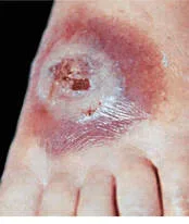
Trypanosomeal chancre at the injection site, painful; stage I illness. 3-5 cm indurated; ulcerate possible. Parasites in fluid expressed from lesion. Fever, lymphadenopathy, arthralgia, malaise, facial edema, and splenomegaly.
The East African variety has a faster course. Arrhythmias and congestive heart failure are also present.
Stage II disease: neurological symptoms.somnolence and indifference.
Melarsoprol, eflorithine, and pentamidine. When treating advanced illness, difluoromethylornithine
Ninety-five percent of reported cases of sleeping sickness in West Africa are caused by Trypanosoma brucei gambiense. East African sleeping sickness is caused by Trypanosoma brucei rhodesiense.
The tse-tse fly is the vector. The main source of West African sleeping disease is humans; antelopes and cattle are the principal carriers of East African sleeping sickness, which affects about 66 million people. Ivory Coast and Chad are in West Africa; laborers in wild regions, rural populations, and tourists are in Central African Republic; and tourism is prevalent in East Africa.
Trypanosomeal chancre at the injection site, painful; stage I illness. 3-5 cm indurated; ulcerate possible. Parasites in fluid expressed from lesion. Fever, lymphadenopathy, arthralgia, malaise, facial edema, and splenomegaly.
The East African variety has a faster course. Arrhythmias and congestive heart failure are also present.
Stage II disease: neurological symptoms.somnolence and indifference.
Melarsoprol, eflorithine, and pentamidine. When treating advanced illness, difluoromethylornithine
- Published on
Dermatology - Leishmaniasis
Sandflies are the primary vector of infection with one of the numerous species of the obligatory intracellular protozoa Leishmania, which causes leishmaniasis.
One to eight weeks after the attack, primary lesions appear at the site of the sandfly bite, usually on the exposed site. There could be lymphadenopathy or lymphangitis.
Skin-colored leishmaniasis At the bite site, a little erythematous papule appears. It develops into an ulcerated nodule that grows to be 3–12 cm in size and has a raised border. Verrucous growths may develop in nonulcerating nodules. Isolated lesions on the head or hand typically heal with a depressed scar and do not ulcerate. Chiclero ulcers are ear sores that can last for years and ruin cartilage. Leprosy-like diffuse cutaneous leishmaniasis is another possibility.
This starts off as a solitary nodule and later spreads locally, frequently via satellite lesion extension, before metastasizing. As the lesions spread throughout the face and trunk, diffuse non-clearing nodules start to form. The sequelae of cutaneous leishmaniasis, known as mucosal leishmaniasis, manifest several years after the initial lesions heal. It is possible for cutaneous and mucosal lesions to occur simultaneously or decades apart.
Epistaxis and coryzal symptoms are caused by edema and inflammatory changes. Gradually, the tonsillar, floor of mouth, and nasal septum are destroyed, leading to noticeable deformity.
Pharyngeal blockage, malnourishment, or an additional bacterial infection can all result in death. One year after treatment, macular, papular, nodular, and hypopigmented macroles/plaques on the face, trunk, and limbs are symptoms of post-Kala-Azar dermal leishmaniasis (PKDL).
By biopsy or culture, the diagnosis is verified. Acute cutaneous leishmaniasis can be differentiated from other conditions such as insect bite reaction, impetigo, ecthyma, furuncle, infection with M. marinum, furuncular myiasis, and chancre.
Meglumine antimoniate and sodium stilbo-gluconate, two substances containing antimony, are administered systemically. Pentamidine, amphotericin B, ketoconazole, miltefosine, paramomycin, and other medications are also used to treat leishmaniasis.
Sandflies are the primary vector of infection with one of the numerous species of the obligatory intracellular protozoa Leishmania, which causes leishmaniasis.
One to eight weeks after the attack, primary lesions appear at the site of the sandfly bite, usually on the exposed site. There could be lymphadenopathy or lymphangitis.
Skin-colored leishmaniasis At the bite site, a little erythematous papule appears. It develops into an ulcerated nodule that grows to be 3–12 cm in size and has a raised border. Verrucous growths may develop in nonulcerating nodules. Isolated lesions on the head or hand typically heal with a depressed scar and do not ulcerate. Chiclero ulcers are ear sores that can last for years and ruin cartilage. Leprosy-like diffuse cutaneous leishmaniasis is another possibility.
This starts off as a solitary nodule and later spreads locally, frequently via satellite lesion extension, before metastasizing. As the lesions spread throughout the face and trunk, diffuse non-clearing nodules start to form. The sequelae of cutaneous leishmaniasis, known as mucosal leishmaniasis, manifest several years after the initial lesions heal. It is possible for cutaneous and mucosal lesions to occur simultaneously or decades apart.
Epistaxis and coryzal symptoms are caused by edema and inflammatory changes. Gradually, the tonsillar, floor of mouth, and nasal septum are destroyed, leading to noticeable deformity.
Pharyngeal blockage, malnourishment, or an additional bacterial infection can all result in death. One year after treatment, macular, papular, nodular, and hypopigmented macroles/plaques on the face, trunk, and limbs are symptoms of post-Kala-Azar dermal leishmaniasis (PKDL).
By biopsy or culture, the diagnosis is verified. Acute cutaneous leishmaniasis can be differentiated from other conditions such as insect bite reaction, impetigo, ecthyma, furuncle, infection with M. marinum, furuncular myiasis, and chancre.
Meglumine antimoniate and sodium stilbo-gluconate, two substances containing antimony, are administered systemically. Pentamidine, amphotericin B, ketoconazole, miltefosine, paramomycin, and other medications are also used to treat leishmaniasis.

- Published on
Dermatology - Diabetic Dermopathy
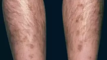
On the anterior lower legs, there are circumscribed, atrophic, somewhat depressed, pink to brown macules that develop in crops and gradually go away. New lesions occasionally develop and may even ulcerate.
A biopsy may be used to confirm the clinical diagnosis, which is made based on the patient's history and physical examination. Necrobiosis lipoidica, pigmented purpuric dermatosis, stasis dermatitis, and pigmented purpuras are among the conditions included in the differential.It is unknown whether or not glucose control is prophylactic, and there is no known effective treatment. Although spontaneous resolution is typical, it is possible for the condition to appear persistent in subsequent crops.
On the anterior lower legs, there are circumscribed, atrophic, somewhat depressed, pink to brown macules that develop in crops and gradually go away. New lesions occasionally develop and may even ulcerate.
A biopsy may be used to confirm the clinical diagnosis, which is made based on the patient's history and physical examination. Necrobiosis lipoidica, pigmented purpuric dermatosis, stasis dermatitis, and pigmented purpuras are among the conditions included in the differential.It is unknown whether or not glucose control is prophylactic, and there is no known effective treatment. Although spontaneous resolution is typical, it is possible for the condition to appear persistent in subsequent crops.