- Published on
Symptoms and Signs – Differential Diagnosis of Bone Pain
• Viral syndrome
• Trauma
• Sickle cell disease
• Paget’s disease of bone
• Osteoporosis with compression fracture
• Osteomalacia
• Neoplasm (primary or metastatic)
• Infection (osteomyelitis, septic arthritis)
• Anxiety
• Viral syndrome
• Trauma
• Sickle cell disease
• Paget’s disease of bone
• Osteoporosis with compression fracture
• Osteomalacia
• Neoplasm (primary or metastatic)
• Infection (osteomyelitis, septic arthritis)
• Anxiety

- Published on
Symptoms and Signs – Differential Diagnosis of Bone Resorption
DISTAL CLAVICLE
• Hyperparathyroidism
• Rheumatoid arthritis (RA)
• Scleroderma
• Post-traumatic osteolysis
• Progeria
• Pycnodysostosis
• Cleidocranial dysplasia
INFERIOR ASPECT OF RIBS
• Vascular impression, associated with but not limited to
coarctation of the aorta
• Hyperparathyroidism
• Neurofibromatosis
TERMINAL PHALANGEAL TUFTS
• Scleroderma
• Raynaud’s phenomenon
• Vascular disease
• Frostbite, electrical burns
• Psoriasis
• Tabes dorsalis
• Hyperparathyroidism
GENERALIZED RESORPTION
• Paraplegia
• Myositis ossificans
• Osteoporosis
DISTAL CLAVICLE
• Hyperparathyroidism
• Rheumatoid arthritis (RA)
• Scleroderma
• Post-traumatic osteolysis
• Progeria
• Pycnodysostosis
• Cleidocranial dysplasia
INFERIOR ASPECT OF RIBS
• Vascular impression, associated with but not limited to
coarctation of the aorta
• Hyperparathyroidism
• Neurofibromatosis
TERMINAL PHALANGEAL TUFTS
• Scleroderma
• Raynaud’s phenomenon
• Vascular disease
• Frostbite, electrical burns
• Psoriasis
• Tabes dorsalis
• Hyperparathyroidism
GENERALIZED RESORPTION
• Paraplegia
• Myositis ossificans
• Osteoporosis
- Published on
Symptoms and Signs – Differential Diagnosis of Primary Malignant Bone Tumor
• Benign bone neoplasm
• Bone cyst
• Metastatic bone disease
• Osteomyelitis
• Benign bone neoplasm
• Bone cyst
• Metastatic bone disease
• Osteomyelitis
- Published on
Symptoms and Signs – Differential Diagnosis of Borderline Personality Disorder
• Substance abuse or dependence: often induces impulsive, emotionally labile behavior.
• Post-traumatic stress disorder (PTSD): individuals with BPD often have history of trauma but do not avoid the feared stimulus or reexperience the trauma, as do individuals with PTSD.
• Mild cases of schizophrenia may superficially resemble BPD.
• Histrionic and narcissistic personality disorders share some common features.
• Dysthymia and other depressive disorders: require a stability of affective symptoms not seen in BPD.
• Bipolar disorder: mood changes in BPD are often triggered by stressors and are less sustained than in bipolar disorder.
• Substance abuse or dependence: often induces impulsive, emotionally labile behavior.
• Post-traumatic stress disorder (PTSD): individuals with BPD often have history of trauma but do not avoid the feared stimulus or reexperience the trauma, as do individuals with PTSD.
• Mild cases of schizophrenia may superficially resemble BPD.
• Histrionic and narcissistic personality disorders share some common features.
• Dysthymia and other depressive disorders: require a stability of affective symptoms not seen in BPD.
• Bipolar disorder: mood changes in BPD are often triggered by stressors and are less sustained than in bipolar disorder.
- Published on
Dermatology - Basal Cell Carcinoma (BCC)
BCC, or basal cell carcinoma, arises from exposure to ultraviolet light and is the most prevalent form of cancer in the human population. BCC exhibits characteristics of local invasiveness, aggressiveness, and destructiveness, while also displaying a moderate growth rate. Metastasis is unlikely to occur unless there is a process of dedifferentiation, such as after receiving insufficient radiation.
However, the tumor has the potential to infiltrate the dura mater, causing destruction of muscle and bone in the brain. BCC typically manifests beyond the age of 40, exhibits a higher prevalence in males, and is infrequent among individuals with dark or black skin.
BCC is a gradually progressing, frequently symptomless abnormality that erodes or bleeds when mildly irritated. Typically, it manifests in the facial region, particularly in the vicinity of the ears and nasolabial folds. The sclerosing kind frequently occurs on the trunk.
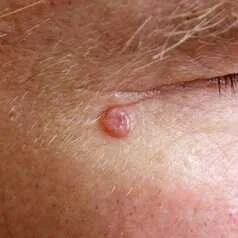
The nodular lesion is characterized by its translucent or pearly appearance, firm texture, and smooth surface. It may also exhibit telangiectasia, and in certain cases, there may be erosions or melanin stippling present. Ulcerating The ulcer is characterized by its translucent appearance, pearly texture, and smooth surface. It often exhibits telangiectasia, and may have a crust and a rolling border. Sclerosing refers to a small area of morphea or superficial scar on the skin. It is typically poorly defined, and can appear flesh-colored, white, or have a speckled pigmentation.
Sclerosing basal cell carcinoma has the potential to develop into either nodular or ulcerating forms. The superficial multicentric form is characterized by thin, pink or red plaques with threadlike edges and telangiectasia. It may also exhibit significant scaling. Superficial multicentric basal cell carcinoma (BCC) can lead to the development of nodular and ulcerating BCC.
Pigmented basal cell carcinoma The lesion can have a smooth, shining surface that ranges in color from brown to blue or black. It is hard, rigid, and typically round or oval in shape. It may also have a cystic or umbilicated appearance.
The diagnosis is made through a clinical assessment that involves a thorough examination with a hand lens, palpation, and dermoscopy. It is then confirmed through histological analysis. The term "differential" encompasses all smooth raised skin lesions. If these lesions are pigmented, they may indicate superficial spreading or nodular melanoma. If the lesions are ulcerated, they may indicate nonpainful hard ulcers such as squamous cell carcinoma or extragenital primary chancre of syphilis. Solar keratoses can be distinguished from BCC by the absence of blood when scraped or irritated.Excisions and repair are considered the standard of care. Cryosurgery and electrosurgery are viable alternatives exclusively for diminutive lesions situated below the neck. Utilize radiation therapy exclusively in cases when surgery may result in notable deformity or in advanced stages of old age. Superficial basal cell carcinomas (BCCs) located below the neck can be effectively treated with cryosurgery or electrocautery with curettage. However, it is important to note that both of these treatment methods can result in scarring. The cream consists of 5-fluorouracil and imiquimod, which are applied directly to the skin. Performing the activity 5 times per week over a period of 6 weeks is both efficient and does not result in scarring. However, it may not completely eliminate all tumor tissue.
BCC, or basal cell carcinoma, arises from exposure to ultraviolet light and is the most prevalent form of cancer in the human population. BCC exhibits characteristics of local invasiveness, aggressiveness, and destructiveness, while also displaying a moderate growth rate. Metastasis is unlikely to occur unless there is a process of dedifferentiation, such as after receiving insufficient radiation.
However, the tumor has the potential to infiltrate the dura mater, causing destruction of muscle and bone in the brain. BCC typically manifests beyond the age of 40, exhibits a higher prevalence in males, and is infrequent among individuals with dark or black skin.
BCC is a gradually progressing, frequently symptomless abnormality that erodes or bleeds when mildly irritated. Typically, it manifests in the facial region, particularly in the vicinity of the ears and nasolabial folds. The sclerosing kind frequently occurs on the trunk.
The nodular lesion is characterized by its translucent or pearly appearance, firm texture, and smooth surface. It may also exhibit telangiectasia, and in certain cases, there may be erosions or melanin stippling present. Ulcerating The ulcer is characterized by its translucent appearance, pearly texture, and smooth surface. It often exhibits telangiectasia, and may have a crust and a rolling border. Sclerosing refers to a small area of morphea or superficial scar on the skin. It is typically poorly defined, and can appear flesh-colored, white, or have a speckled pigmentation.
Sclerosing basal cell carcinoma has the potential to develop into either nodular or ulcerating forms. The superficial multicentric form is characterized by thin, pink or red plaques with threadlike edges and telangiectasia. It may also exhibit significant scaling. Superficial multicentric basal cell carcinoma (BCC) can lead to the development of nodular and ulcerating BCC.
Pigmented basal cell carcinoma The lesion can have a smooth, shining surface that ranges in color from brown to blue or black. It is hard, rigid, and typically round or oval in shape. It may also have a cystic or umbilicated appearance.
The diagnosis is made through a clinical assessment that involves a thorough examination with a hand lens, palpation, and dermoscopy. It is then confirmed through histological analysis. The term "differential" encompasses all smooth raised skin lesions. If these lesions are pigmented, they may indicate superficial spreading or nodular melanoma. If the lesions are ulcerated, they may indicate nonpainful hard ulcers such as squamous cell carcinoma or extragenital primary chancre of syphilis. Solar keratoses can be distinguished from BCC by the absence of blood when scraped or irritated.Excisions and repair are considered the standard of care. Cryosurgery and electrosurgery are viable alternatives exclusively for diminutive lesions situated below the neck. Utilize radiation therapy exclusively in cases when surgery may result in notable deformity or in advanced stages of old age. Superficial basal cell carcinomas (BCCs) located below the neck can be effectively treated with cryosurgery or electrocautery with curettage. However, it is important to note that both of these treatment methods can result in scarring. The cream consists of 5-fluorouracil and imiquimod, which are applied directly to the skin. Performing the activity 5 times per week over a period of 6 weeks is both efficient and does not result in scarring. However, it may not completely eliminate all tumor tissue.
- Published on
Dermatology - Acne
Acne is a prevalent condition that affects around 85% of young individuals. It usually starts during puberty, although it can also manifest for the first time at the age of 25 or older. Acne has greater severity in males compared to females and is less common among individuals of Asian and African ancestry. The presence of multiple genetic factors and a familial propensity contribute to this condition. The majority of persons with cystic acne have parents who have a history of severe acne.
Lesions, particularly of the nodulocystic variety, can cause pain and persist for several weeks to months. They tend to worsen throughout the autumn and winter seasons. Lesions manifest on the face, neck, trunk, upper arms, and buttocks.
Abnormalities
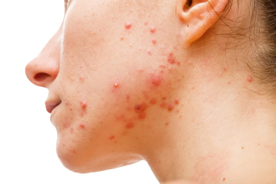
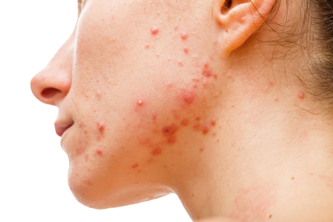
Comedones refer to two types of acne: open comedones, commonly known as blackheads, and closed comedones, also known as whiteheads. They are both forms of comedonal acne. Papules and papulopustules refer to skin lesions characterized by the presence of a raised bump (papule) that is topped with a small blister filled with pus (pustule). This condition is commonly known as papulopustular acne. Nodules or cysts are often 1-4 cm in diameter. Nodulocystic acne occurs when soft nodules form due to frequent follicular ruptures and subsequent reencapsulations accompanied by inflammation, abscess formation (cysts), and foreign-body reaction.
Comedones are necessary for diagnosing any kind of acne and are not present in acne-like conditions or the following conditions: Facial conditions include S. aureus folliculitis, pseudofolliculitis barbae, rosacea, and perioral dermatitis. Trunk: Inflammation of hair follicles caused by Malassezia, pseudomonas infection from hot tubs, or Staphylococcus aureus infection.
Mild acne
Apply topical antibiotics such as clindamycin and erythromycin, as well as benzoyl peroxide gels with concentrations of 2%, 5%, or 10%. Topical retinoids can be utilized by following specific guidelines that involve gradually increasing the concentration of cream/gel or liquid from 0.01% to 0.025% to 0.05%. They can also be combined with benzoyl peroxide-erythromycin gels. Extraction is only beneficial when performed correctly following pretreatment with topical retinoids.
Mild to moderate acne
Incorporate oral antibiotics into the aforementioned treatment plan. Minocycline, administered at a dosage of 50-100 mg twice day, or doxycycline, also at a dosage of 50-100 mg twice daily, can be highly beneficial. The dosage of doxycycline can be gradually reduced to 50 mg per day as the severity of acne decreases.
Oral administration of isotretinoin is efficacious in preventing scarring.
Severe acne
Administer isotretinoin as a systemic treatment for cystic or conglobate acne, as well as for any other forms of acne that do not respond to other treatments.
Acne is a prevalent condition that affects around 85% of young individuals. It usually starts during puberty, although it can also manifest for the first time at the age of 25 or older. Acne has greater severity in males compared to females and is less common among individuals of Asian and African ancestry. The presence of multiple genetic factors and a familial propensity contribute to this condition. The majority of persons with cystic acne have parents who have a history of severe acne.
Lesions, particularly of the nodulocystic variety, can cause pain and persist for several weeks to months. They tend to worsen throughout the autumn and winter seasons. Lesions manifest on the face, neck, trunk, upper arms, and buttocks.
Abnormalities
Comedones refer to two types of acne: open comedones, commonly known as blackheads, and closed comedones, also known as whiteheads. They are both forms of comedonal acne. Papules and papulopustules refer to skin lesions characterized by the presence of a raised bump (papule) that is topped with a small blister filled with pus (pustule). This condition is commonly known as papulopustular acne. Nodules or cysts are often 1-4 cm in diameter. Nodulocystic acne occurs when soft nodules form due to frequent follicular ruptures and subsequent reencapsulations accompanied by inflammation, abscess formation (cysts), and foreign-body reaction.
Comedones are necessary for diagnosing any kind of acne and are not present in acne-like conditions or the following conditions: Facial conditions include S. aureus folliculitis, pseudofolliculitis barbae, rosacea, and perioral dermatitis. Trunk: Inflammation of hair follicles caused by Malassezia, pseudomonas infection from hot tubs, or Staphylococcus aureus infection.
Mild acne
Apply topical antibiotics such as clindamycin and erythromycin, as well as benzoyl peroxide gels with concentrations of 2%, 5%, or 10%. Topical retinoids can be utilized by following specific guidelines that involve gradually increasing the concentration of cream/gel or liquid from 0.01% to 0.025% to 0.05%. They can also be combined with benzoyl peroxide-erythromycin gels. Extraction is only beneficial when performed correctly following pretreatment with topical retinoids.
Mild to moderate acne
Incorporate oral antibiotics into the aforementioned treatment plan. Minocycline, administered at a dosage of 50-100 mg twice day, or doxycycline, also at a dosage of 50-100 mg twice daily, can be highly beneficial. The dosage of doxycycline can be gradually reduced to 50 mg per day as the severity of acne decreases.
Oral administration of isotretinoin is efficacious in preventing scarring.
Severe acne
Administer isotretinoin as a systemic treatment for cystic or conglobate acne, as well as for any other forms of acne that do not respond to other treatments.
- Published on
Dermatology - Granuloma Annulare
Granuloma annulare is a frequently occurring, self-limiting, asymptomatic, and persistent skin condition that typically affects children and young adults.
Granuloma annulare has a duration of many months to years and typically does not cause any symptoms, except for cosmetic damage.
Abnormalities
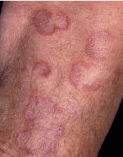
The individual presents with raised, even, glossy, clustered skin lesions and patches, measuring 1-5 cm in diameter. These lesions have a circular or crescent shape with a depressed center and can appear in various colors, including flesh-colored, bluish-purple, or red.
Subcutaneous granuloma annulare is an uncommon and painless condition characterized by the presence of skin-colored nodules that develop in the deep layers of the skin or just beneath it. These nodules can arise either as a single nodule or as many nodules, and are typically found on the fingers and toes. The lesions are primarily found on the back of the hand, fingers, or lower arm. Multiple lesions can manifest on the limbs and torso, or be widespread in nature (papular; in elderly individuals). Subcutaneous lesions are situated in close proximity to joints, palms, soles, and buttocks.
The diagnosis is made based on clinical assessment after excluding more severe disorders. Precise diagnosis is crucial due to its resemblance to more severe illnesses. The differential diagnosis comprises necrobiosis lipoidica, papular sarcoid, lichen planus, lymphocytic infiltration of Jessner, rheumatoid nodules, and subcutaneous fungal infections such as sporotrichosis, tinea, erythema migrans, and lichen planus.
Granuloma annulare is a localized skin illness that does not indicate the presence of an interior disease. Spontaneous remission is typically observed. If the lesions are not causing any disfigurement, then it is possible to consider not pursuing any therapy.
Lesions may heal after undergoing a biopsy procedure. If treatment is sought for cosmetic purposes, the application of glucocorticoids on the skin with occlusion or the injection of triamcinolone at a concentration of 3 mg/mL directly into the affected area are both effective methods.
Cryotherapy with liquid nitrogen is effective in treating superficial lesions, although it may lead to atrophy.
Photodynamic treatment is efficacious for widespread lesions. Systemic glucocorticoids are also efficacious in treating widespread lesions, but recurrences are frequent.
Granuloma annulare is a frequently occurring, self-limiting, asymptomatic, and persistent skin condition that typically affects children and young adults.
Granuloma annulare has a duration of many months to years and typically does not cause any symptoms, except for cosmetic damage.
Abnormalities
The individual presents with raised, even, glossy, clustered skin lesions and patches, measuring 1-5 cm in diameter. These lesions have a circular or crescent shape with a depressed center and can appear in various colors, including flesh-colored, bluish-purple, or red.
Subcutaneous granuloma annulare is an uncommon and painless condition characterized by the presence of skin-colored nodules that develop in the deep layers of the skin or just beneath it. These nodules can arise either as a single nodule or as many nodules, and are typically found on the fingers and toes. The lesions are primarily found on the back of the hand, fingers, or lower arm. Multiple lesions can manifest on the limbs and torso, or be widespread in nature (papular; in elderly individuals). Subcutaneous lesions are situated in close proximity to joints, palms, soles, and buttocks.
The diagnosis is made based on clinical assessment after excluding more severe disorders. Precise diagnosis is crucial due to its resemblance to more severe illnesses. The differential diagnosis comprises necrobiosis lipoidica, papular sarcoid, lichen planus, lymphocytic infiltration of Jessner, rheumatoid nodules, and subcutaneous fungal infections such as sporotrichosis, tinea, erythema migrans, and lichen planus.
Granuloma annulare is a localized skin illness that does not indicate the presence of an interior disease. Spontaneous remission is typically observed. If the lesions are not causing any disfigurement, then it is possible to consider not pursuing any therapy.
Lesions may heal after undergoing a biopsy procedure. If treatment is sought for cosmetic purposes, the application of glucocorticoids on the skin with occlusion or the injection of triamcinolone at a concentration of 3 mg/mL directly into the affected area are both effective methods.
Cryotherapy with liquid nitrogen is effective in treating superficial lesions, although it may lead to atrophy.
Photodynamic treatment is efficacious for widespread lesions. Systemic glucocorticoids are also efficacious in treating widespread lesions, but recurrences are frequent.
- Published on
Dermatology - Scleroderma
Inflammatory, vascular, and sclerotic alterations of the skin and several internal organs, including the heart, lungs, and gastrointestinal tract, are the hallmarks of scleroderma, a multisystem condition.
60% of instances with scleroderma are restricted, and 40% are diffuse; the latter has a high rate of morbidity and death. It strikes between the ages of 30 and 50, with women four times more likely to be impacted than males.
There is always secondary Raynaud phenomenon and skin sclerosis. Additional symptoms include heartburn, migratory polyarthritis, arthralgia, constipation, diarrhea, bloating in the abdomen, malabsorption, weight loss, exertional dyspnea, and dry cough.
The Raynaud phenomenon occurs months or years before sclerosis. Next, there is painful ulcerations at the fingertips accompanied by nonpitting edema of the hands and feet that heals with pitted scars. Later on, sclerodactyly develops, characterized by tapering fingers and skin that is glossy, waxy, and rigid, tightly bound to prevent folding or wrinkles. Synovial flexion contractures cause leathery crepitation over those joints. Nails with shorter distal phalanges become claw-like due to periungual telangiectasia. Distal phalanges are lost as a consequence of resorption of bone and ulcerations.
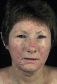
On the distal extremities, there is total hair loss, thinning, and anhidrosis associated with the loss of sweat glands. Periorbital edema is the first symptom, and more edema and fibrosis cause mask-like facies, radial perioral furrowing, thinning of the lips, beak-like pointed nose, and loss of normal facial lines. There is diffuse hyperpigmentation and telangiectasia. Diffuse scleroderma is characterized by early involvement of the chest and proximal upper and lower extremities, as well as tense, stiff, and waxy-looking skin that is impossible to fold and limited chest wall movement, which impairs breathing.
Clinical findings are used to make the diagnosis, which is then verified by dermatopathology. Mixed connective tissue disease, eosinophilic fasciitis, scleromyxedema, morphea, porphyria cutanea tarda, adverse drug responses (bleomycin, pentazocine), polyvinyl chloride exposure, lichen sclerosus et atrophicus, and systemic fibrosis are among the conditions included in the differential.
Early in the disease, systemic glucocorticoids may be helpful for brief periods of time.
Immunosuppressive medications such as mycophenolate mofetil, cyclosporine, methotrexate, and cyclophosphamide have improved skin, but their effects on systemic involvement have been somewhat restricted.
Inflammatory, vascular, and sclerotic alterations of the skin and several internal organs, including the heart, lungs, and gastrointestinal tract, are the hallmarks of scleroderma, a multisystem condition.
60% of instances with scleroderma are restricted, and 40% are diffuse; the latter has a high rate of morbidity and death. It strikes between the ages of 30 and 50, with women four times more likely to be impacted than males.
There is always secondary Raynaud phenomenon and skin sclerosis. Additional symptoms include heartburn, migratory polyarthritis, arthralgia, constipation, diarrhea, bloating in the abdomen, malabsorption, weight loss, exertional dyspnea, and dry cough.
The Raynaud phenomenon occurs months or years before sclerosis. Next, there is painful ulcerations at the fingertips accompanied by nonpitting edema of the hands and feet that heals with pitted scars. Later on, sclerodactyly develops, characterized by tapering fingers and skin that is glossy, waxy, and rigid, tightly bound to prevent folding or wrinkles. Synovial flexion contractures cause leathery crepitation over those joints. Nails with shorter distal phalanges become claw-like due to periungual telangiectasia. Distal phalanges are lost as a consequence of resorption of bone and ulcerations.
On the distal extremities, there is total hair loss, thinning, and anhidrosis associated with the loss of sweat glands. Periorbital edema is the first symptom, and more edema and fibrosis cause mask-like facies, radial perioral furrowing, thinning of the lips, beak-like pointed nose, and loss of normal facial lines. There is diffuse hyperpigmentation and telangiectasia. Diffuse scleroderma is characterized by early involvement of the chest and proximal upper and lower extremities, as well as tense, stiff, and waxy-looking skin that is impossible to fold and limited chest wall movement, which impairs breathing.
Clinical findings are used to make the diagnosis, which is then verified by dermatopathology. Mixed connective tissue disease, eosinophilic fasciitis, scleromyxedema, morphea, porphyria cutanea tarda, adverse drug responses (bleomycin, pentazocine), polyvinyl chloride exposure, lichen sclerosus et atrophicus, and systemic fibrosis are among the conditions included in the differential.
Early in the disease, systemic glucocorticoids may be helpful for brief periods of time.
Immunosuppressive medications such as mycophenolate mofetil, cyclosporine, methotrexate, and cyclophosphamide have improved skin, but their effects on systemic involvement have been somewhat restricted.
- Published on
Dermatology - Mongolian Spots
Congenital pigmented lesions are known as Mongolian spots.
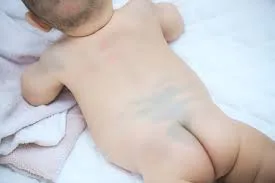
Gray-blue macular lesions can appear anywhere on the skin, including the back, scalp, and lumbosacral region, where they are typically found. Typically, there is only one lesion; but, in rare occasions, a newborn may have many truncal lesions.Clinical diagnosis is made.
Other than reassuring parents, no treatment is necessary for these lesions as no melanomas have been documented to develop in them.
Congenital pigmented lesions are known as Mongolian spots.
Gray-blue macular lesions can appear anywhere on the skin, including the back, scalp, and lumbosacral region, where they are typically found. Typically, there is only one lesion; but, in rare occasions, a newborn may have many truncal lesions.Clinical diagnosis is made.
Other than reassuring parents, no treatment is necessary for these lesions as no melanomas have been documented to develop in them.
- Published on
Dermatology - Lipoma
The fat cells that make up lipomas are normal-looking fat cells enclosed in a framework of connective tissue. Vascular elements are present in angiolipomas. Hundreds of slowly growing, nontender lesions are the hallmark of the autosomal dominant feature known as familial lipoma syndrome, which first manifests in early adulthood. Middle-aged women who develop adipositas dolorosa, also known as Dercum disease, have numerous painful, widespread fatty deposits that are not confined. Men in their middle age have benign symmetric lipomatosis, which is characterized by numerous big, nontender, coalescent, poorly delimited lipomas, primarily on the trunk and upper extremities; these lipomas can combine around the neck and give the impression of a "horse-collar."
Angiolipomas may become sensitive when compressed and in a chilly environment.
Damage
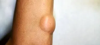
Lipomas are benign subcutaneous tumors that can range in size from one to several and up to more than six centimeters. They can be moved in relation to the skin above and are pliable, rounded, or lobulated.
Although they can develop anywhere, lipomas typically affect the neck, trunk, and extremities.
Clinical diagnosis is made.
While other lipomas should only be removed if they are thought to be disfiguring, angiolipomas frequently need to be removed. When lipomas are soft and have little to no connective tissue, liposuction can also be used.
The fat cells that make up lipomas are normal-looking fat cells enclosed in a framework of connective tissue. Vascular elements are present in angiolipomas. Hundreds of slowly growing, nontender lesions are the hallmark of the autosomal dominant feature known as familial lipoma syndrome, which first manifests in early adulthood. Middle-aged women who develop adipositas dolorosa, also known as Dercum disease, have numerous painful, widespread fatty deposits that are not confined. Men in their middle age have benign symmetric lipomatosis, which is characterized by numerous big, nontender, coalescent, poorly delimited lipomas, primarily on the trunk and upper extremities; these lipomas can combine around the neck and give the impression of a "horse-collar."
Angiolipomas may become sensitive when compressed and in a chilly environment.
Damage
Lipomas are benign subcutaneous tumors that can range in size from one to several and up to more than six centimeters. They can be moved in relation to the skin above and are pliable, rounded, or lobulated.
Although they can develop anywhere, lipomas typically affect the neck, trunk, and extremities.
Clinical diagnosis is made.
While other lipomas should only be removed if they are thought to be disfiguring, angiolipomas frequently need to be removed. When lipomas are soft and have little to no connective tissue, liposuction can also be used.