- Published on
Dermatology - Impetigo
The primary causative agents of impetigo are typically Staphylococcus aureus or beta-hemolytic streptococcus group A. Bullous impetigo occurs due to the localized synthesis of epidermolytic toxin by S. aureus bacteria. These microorganisms are not commonly found on human skin, but instead temporarily inhabit the skin and produce surface-level diseases. Primary infections generally manifest in youngsters, although both primary and secondary infections can occur across all age groups.
Superficial infections frequently lack noticeable symptoms.
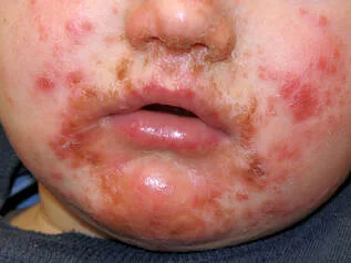
Impetigo manifests as erosions characterized by golden-yellow crusts measuring 1 to over 3 cm in diameter. These lesions exhibit central healing after several weeks of presence. The lesions are dispersed, distinct, and can merge together; satellite lesions may appear due to self-inoculation. Secondary infection frequently occurs. Bullous Impetigo manifests as blisters filled with translucent yellow or slightly cloudy fluid, surrounded by a red ring, on skin that appears normal. Rupture leads to the decompression of bullous lesions. Removing the roof of a bulla leads to the development of a shallow and damp erosion.
Ecthyma is a condition characterized by the formation of ulcers covered by a thick, firmly attached crust. These ulcers are often painful and hardened.
The diagnosis is made based on clinical observations and validated with laboratory culture tests. The differential diagnosis comprises excoriation, allergic contact dermatitis, herpes simplex, epidermal dermatophytosis, scabies, burns, porphyria cutanea tarda, venous stasis, and ischemic ulcers.Manage skin sores using mupirocin and retapamulin ointment, and avoid future occurrences by using benzoyl peroxide wash and/or applying mupirocin and retapamulin ointment to the nostrils.
Examine family members for symptoms of impetigo and urge all individuals in close proximity to cleanse their hands with ethanol or isopropyl gel.
The primary causative agents of impetigo are typically Staphylococcus aureus or beta-hemolytic streptococcus group A. Bullous impetigo occurs due to the localized synthesis of epidermolytic toxin by S. aureus bacteria. These microorganisms are not commonly found on human skin, but instead temporarily inhabit the skin and produce surface-level diseases. Primary infections generally manifest in youngsters, although both primary and secondary infections can occur across all age groups.
Superficial infections frequently lack noticeable symptoms.
Impetigo manifests as erosions characterized by golden-yellow crusts measuring 1 to over 3 cm in diameter. These lesions exhibit central healing after several weeks of presence. The lesions are dispersed, distinct, and can merge together; satellite lesions may appear due to self-inoculation. Secondary infection frequently occurs. Bullous Impetigo manifests as blisters filled with translucent yellow or slightly cloudy fluid, surrounded by a red ring, on skin that appears normal. Rupture leads to the decompression of bullous lesions. Removing the roof of a bulla leads to the development of a shallow and damp erosion.
Ecthyma is a condition characterized by the formation of ulcers covered by a thick, firmly attached crust. These ulcers are often painful and hardened.
The diagnosis is made based on clinical observations and validated with laboratory culture tests. The differential diagnosis comprises excoriation, allergic contact dermatitis, herpes simplex, epidermal dermatophytosis, scabies, burns, porphyria cutanea tarda, venous stasis, and ischemic ulcers.Manage skin sores using mupirocin and retapamulin ointment, and avoid future occurrences by using benzoyl peroxide wash and/or applying mupirocin and retapamulin ointment to the nostrils.
Examine family members for symptoms of impetigo and urge all individuals in close proximity to cleanse their hands with ethanol or isopropyl gel.
- Published on
Dermatology - Beau Lines
Beau lines
Beau lines occur when nail development is temporarily halted following a severe, abrupt, acute sickness, such as a medication reaction or in the presence of atopy, peripheral vasculitis, or ischemia. If all 20 nails are affected, it suggests the presence of a systemic disease. Typically, single nail involvement is the result of a stressful event.
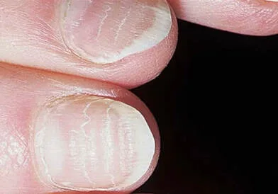
The nails are affected by transverse depressions that reach from one side to the other, affecting all nails at the same level. If the length of the disease entirely halts the activity of the matrix for a period of 7-14 days, it leads to the formation of a transverse depression that causes the nail plate to totally separate (a condition known as onychomadesis). Chemotherapy can involve the use of numerous parallel lines.
Beau lines do not constitute a standalone diagnosis, but rather indicate an underlying or recent disease.
Beau lines will gradually go as the nail regenerates and any underlying ailment is addressed.
No specific management is needed.
Beau lines
Beau lines occur when nail development is temporarily halted following a severe, abrupt, acute sickness, such as a medication reaction or in the presence of atopy, peripheral vasculitis, or ischemia. If all 20 nails are affected, it suggests the presence of a systemic disease. Typically, single nail involvement is the result of a stressful event.
The nails are affected by transverse depressions that reach from one side to the other, affecting all nails at the same level. If the length of the disease entirely halts the activity of the matrix for a period of 7-14 days, it leads to the formation of a transverse depression that causes the nail plate to totally separate (a condition known as onychomadesis). Chemotherapy can involve the use of numerous parallel lines.
Beau lines do not constitute a standalone diagnosis, but rather indicate an underlying or recent disease.
Beau lines will gradually go as the nail regenerates and any underlying ailment is addressed.
No specific management is needed.
- Published on
Dermatology - Lyme Disease
Lyme disease, which is caused by spirochetes called Borrelia, is the most prevalent vector-borne illness in the US, accounting for 30,000 cases annually. The disease is spread via tick bites.
Stage 1: Up to 30 days after a tick bite, a severe sickness with fever, chills, myalgia, headache, weakness, and photophobia might ensue. Stage 2: Symptoms that appear days to weeks after the bite include neurologic (meningitis, cranial neuritis, and peripheral neuritis), cardiac (AV nodal block), and musculoskeletal (myalgia, arthralgia). Stage 3: Weeks to months later, polyneuropathy, chronic arthralgia, and persistent arthritis develop.
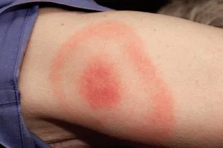
Stage 1: An erythematous plaque, also known as erythema migrans, characterized by a red border and a fast centrifugal lesion expansion. The lesion may either produce multiple concentric rings of varied shades of red (targetoid look) or remain uniformly red as it grows.
Days to weeks after a tick bite, stage 2 happens. Similar lesions to erythema migrants are observed; however, they differ in size, travel less, lack central induration, and sometimes have scaly appearance.
Stage 3 happens when acrodermatitis chronica atrophicans first appears, it usually affects one extremity and is characterized by a diffuse or localized violaceous erythema, modest to substantial edema, and Lyme disease. Over months to years, the lesion spreads centrifugally, leaving behind visible veins, subcutaneous tissue, and central atrophy. Subcutaneous nodules around the knees and elbows are indicative of localized fibromas and plaques.
For Stage 1 disease, the diagnosis is made clinically; for Stages 2 and 3, the diagnosis is determined based on the presence of antibodies. Other insect bites, tinea, allergic contact dermatitis, pityriasis rosea, fixed drug eruption, erythema multiforme, and urticaria are among the differential diagnoses.
Doxycycline, 100 mg twice day, is the first-line treatment for all patients over 9 years old who are not pregnant. Give amoxicillin 50 mg/kg daily to children younger than nine years old.
For adults, amoxicillin 500 mg three times a day is the second-line treatment. Erythromycin 250 mg four times a day or cefuroxime axetil 500 mg twice a day are substitutes.
For Stage 1 disease, continue antibiotics for 14 days; for Stage 2 disease, continue for 21 days; and for Stage 3 disease, continue for 30–60 days. For any patients exhibiting cardiac or neurological symptoms, think about administering IV antibiotics.
Lyme disease, which is caused by spirochetes called Borrelia, is the most prevalent vector-borne illness in the US, accounting for 30,000 cases annually. The disease is spread via tick bites.
Stage 1: Up to 30 days after a tick bite, a severe sickness with fever, chills, myalgia, headache, weakness, and photophobia might ensue. Stage 2: Symptoms that appear days to weeks after the bite include neurologic (meningitis, cranial neuritis, and peripheral neuritis), cardiac (AV nodal block), and musculoskeletal (myalgia, arthralgia). Stage 3: Weeks to months later, polyneuropathy, chronic arthralgia, and persistent arthritis develop.
Stage 1: An erythematous plaque, also known as erythema migrans, characterized by a red border and a fast centrifugal lesion expansion. The lesion may either produce multiple concentric rings of varied shades of red (targetoid look) or remain uniformly red as it grows.
Days to weeks after a tick bite, stage 2 happens. Similar lesions to erythema migrants are observed; however, they differ in size, travel less, lack central induration, and sometimes have scaly appearance.
Stage 3 happens when acrodermatitis chronica atrophicans first appears, it usually affects one extremity and is characterized by a diffuse or localized violaceous erythema, modest to substantial edema, and Lyme disease. Over months to years, the lesion spreads centrifugally, leaving behind visible veins, subcutaneous tissue, and central atrophy. Subcutaneous nodules around the knees and elbows are indicative of localized fibromas and plaques.
For Stage 1 disease, the diagnosis is made clinically; for Stages 2 and 3, the diagnosis is determined based on the presence of antibodies. Other insect bites, tinea, allergic contact dermatitis, pityriasis rosea, fixed drug eruption, erythema multiforme, and urticaria are among the differential diagnoses.
Doxycycline, 100 mg twice day, is the first-line treatment for all patients over 9 years old who are not pregnant. Give amoxicillin 50 mg/kg daily to children younger than nine years old.
For adults, amoxicillin 500 mg three times a day is the second-line treatment. Erythromycin 250 mg four times a day or cefuroxime axetil 500 mg twice a day are substitutes.
For Stage 1 disease, continue antibiotics for 14 days; for Stage 2 disease, continue for 21 days; and for Stage 3 disease, continue for 30–60 days. For any patients exhibiting cardiac or neurological symptoms, think about administering IV antibiotics.
- Published on
Dermatology - Tinea Cruris
A particular class of fungus known as dermatiophytes is responsible for the infection of keratinized cutaneous tissues that causes tinea; arthrospores from these species can live for up to a year in skin scales.
The most prevalent ways for transmission are from person to person, from animals, and, less frequently, via soil. Tinea cruris is a subacute or chronic dermatophytosis that affects the pubic and inguinal regions, as well as the upper thigh. It is nearly invariably linked to tinea pedis.
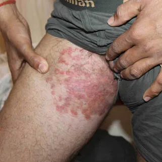
Erythematous macules of approximately 3-5 mm and either scaling or not combine to create big, well-defined, dull red, tan, or brown plaques with a central clearing. Dermatophytic folliculitis and pustules and papules may be seen at the edges. Lichen simplex chronicus subsequent alterations may result from persistent scratching. Seldom are the penis and scrotum implicated.
Fungal hyphae can be seen by direct microscopy of skin scrapings that have been taken using a no. 15 scalpel blade, the edge of a glass microscope slide, or a toothbrush (cervical or tooth brush) and covered with a drop of potassium hydroxide (KOH). Erythasma, candida intertrigo, intertriginous psoriasis, tinea, or pityriasis versicolor are among the conditions included in the differential.
Use imidazole powder, benzoyl peroxide wash, and dry skin to stop recurrences. If dermatophytic nail infection is present, make careful to treat it.
Subject-specific
Use allylamine lotion (naftifin, terbinafine); naphthionate ointment (tolnaftate); imidazole creams (clotrimazole, miconazole, ketoconazole, econazole, oxiconizole, sudonizole, sertaconazole); or substituted pyridine (ciclopirox olamine).
Among oral antidermophytic agents, Systemic Terbinafine 250-mg tablet is the most effective allylamine. Fluconazole 100-, 150-, and 200-mg pills or oral suspension (10 or 40 mg/mL) are substitutes for itraconazole 100-mg capsules or oral solution (10 mg/mL).
A particular class of fungus known as dermatiophytes is responsible for the infection of keratinized cutaneous tissues that causes tinea; arthrospores from these species can live for up to a year in skin scales.
The most prevalent ways for transmission are from person to person, from animals, and, less frequently, via soil. Tinea cruris is a subacute or chronic dermatophytosis that affects the pubic and inguinal regions, as well as the upper thigh. It is nearly invariably linked to tinea pedis.
Erythematous macules of approximately 3-5 mm and either scaling or not combine to create big, well-defined, dull red, tan, or brown plaques with a central clearing. Dermatophytic folliculitis and pustules and papules may be seen at the edges. Lichen simplex chronicus subsequent alterations may result from persistent scratching. Seldom are the penis and scrotum implicated.
Fungal hyphae can be seen by direct microscopy of skin scrapings that have been taken using a no. 15 scalpel blade, the edge of a glass microscope slide, or a toothbrush (cervical or tooth brush) and covered with a drop of potassium hydroxide (KOH). Erythasma, candida intertrigo, intertriginous psoriasis, tinea, or pityriasis versicolor are among the conditions included in the differential.
Use imidazole powder, benzoyl peroxide wash, and dry skin to stop recurrences. If dermatophytic nail infection is present, make careful to treat it.
Subject-specific
Use allylamine lotion (naftifin, terbinafine); naphthionate ointment (tolnaftate); imidazole creams (clotrimazole, miconazole, ketoconazole, econazole, oxiconizole, sudonizole, sertaconazole); or substituted pyridine (ciclopirox olamine).
Among oral antidermophytic agents, Systemic Terbinafine 250-mg tablet is the most effective allylamine. Fluconazole 100-, 150-, and 200-mg pills or oral suspension (10 or 40 mg/mL) are substitutes for itraconazole 100-mg capsules or oral solution (10 mg/mL).
- Published on
Dermatology - Migratory Glossitis
Geographic tongue, also known as migratory glossitis, is a widespread condition with an unclear cause.
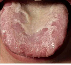
Elevated white or yellow edges encircle irregular regions of red, dekeratinized, and desquamated filiform papillae margins.
Clinical diagnosis is made.
There is no need for management because this is a common, benign illness.
Geographic tongue, also known as migratory glossitis, is a widespread condition with an unclear cause.
Elevated white or yellow edges encircle irregular regions of red, dekeratinized, and desquamated filiform papillae margins.
Clinical diagnosis is made.
There is no need for management because this is a common, benign illness.
- Published on
Dermatology - Sarcoidosis
A systemic granulomatous illness with a range of cutaneous manifestations, sarcoidosis primarily affects the lungs (bilateral lymphadenopathy, pulmonary infiltration) and has an unknown etiology. Onset often occurs before 40.
Fever, exhaustion, weight loss, and arrhythmia are examples of constitutional symptoms. Lesions might appear days after other symptoms (acute erythema nodosum) or months later (asymptomatic sarcoidal papules, plaques on the skin, or pulmonary infiltration seen on regular chest radiography).
The skin-colored papules around the mouth are the first lesions to appear. Afterwards, the trunk, buttocks, and extremities are the primary sites for brownish or purple infiltrating plaques that can be annular, polycyclic, or serpiginous. There may be central clearing accompanied by mild atrophy. Firm purple or brown nodules on the face, torso, or extremities—especially the hands—may occasionally appear.
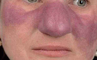
Diffuse, violaceous, soft, doughy infiltrations on the nose, cheekbones, or earlobes are indicative of lupus pernio.
Because of osteitis cystica, individual digits may enlarge. Old scars are often infiltrated by sarcoidosis, resulting in translucent purple-red or yellowish papules or nodules. All cutaneous lesions of sarcoidosis exhibit a semitranslucent yellowish brown tint known as "apple jelly" when blanched on a glass slide. Sarcoidosis can result in scarring alopecia on the scalp.
Skin or lymph node lesional biopsies are used to make the diagnosis.
When there is a CNS involvement, cardiac arrhythmia, active lung disease, active ocular disease, or hypercalcemia, systemic glucocorticoids are used. Intralesional triamcinolone (3 mg/mL) is useful for minor lesions in cutaneous sarcoidosis, but systemic glucocorticoids are administered for extensive or deformity-causing involvement. When intralesional triamcinolone is ineffective for widespread or disfiguring lesions, low-dose methotrexate or 100 mg of hydroxychloroquine twice day may be administered; however, these treatments are not always successful.
Only diseases that have the potential to be fatal are treated with cyclophosphamide.
A systemic granulomatous illness with a range of cutaneous manifestations, sarcoidosis primarily affects the lungs (bilateral lymphadenopathy, pulmonary infiltration) and has an unknown etiology. Onset often occurs before 40.
Fever, exhaustion, weight loss, and arrhythmia are examples of constitutional symptoms. Lesions might appear days after other symptoms (acute erythema nodosum) or months later (asymptomatic sarcoidal papules, plaques on the skin, or pulmonary infiltration seen on regular chest radiography).
The skin-colored papules around the mouth are the first lesions to appear. Afterwards, the trunk, buttocks, and extremities are the primary sites for brownish or purple infiltrating plaques that can be annular, polycyclic, or serpiginous. There may be central clearing accompanied by mild atrophy. Firm purple or brown nodules on the face, torso, or extremities—especially the hands—may occasionally appear.
Diffuse, violaceous, soft, doughy infiltrations on the nose, cheekbones, or earlobes are indicative of lupus pernio.
Because of osteitis cystica, individual digits may enlarge. Old scars are often infiltrated by sarcoidosis, resulting in translucent purple-red or yellowish papules or nodules. All cutaneous lesions of sarcoidosis exhibit a semitranslucent yellowish brown tint known as "apple jelly" when blanched on a glass slide. Sarcoidosis can result in scarring alopecia on the scalp.
Skin or lymph node lesional biopsies are used to make the diagnosis.
When there is a CNS involvement, cardiac arrhythmia, active lung disease, active ocular disease, or hypercalcemia, systemic glucocorticoids are used. Intralesional triamcinolone (3 mg/mL) is useful for minor lesions in cutaneous sarcoidosis, but systemic glucocorticoids are administered for extensive or deformity-causing involvement. When intralesional triamcinolone is ineffective for widespread or disfiguring lesions, low-dose methotrexate or 100 mg of hydroxychloroquine twice day may be administered; however, these treatments are not always successful.
Only diseases that have the potential to be fatal are treated with cyclophosphamide.
- Published on
Dermatology - Squamous Cell Carcinoma (SCCIS)
HPV infection or UV radiation are the main causes of SCCIS. It frequently manifests itself in dysplastic lesions like solar keratoses. Bowen disease, erythroplasia (onglans/vulva), and anogenital, HPV-induced bowenoid papulosis are among the synonyms. Invasive squamous cell carcinoma (SCC) can develop from SSCIS. Without obvious invasion, lymph node metastasis can happen and spread.
One or more hyperkeratotic or scaling macules, papules, or plaques that may bleed in addition to being typically asymptomatic. Nodule formation, discomfort, or soreness indicate that SCC is progressing.
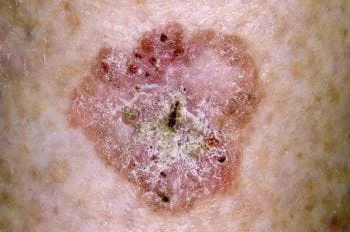
Sharply defined, scaling, or hyperkeratotic pink or red macules, papules, or plaques characterize the lesions. Lesions can be crusty and might be single or numerous. Scaling or crusting lesions, whether single or many, are consistently well-defined (Bowen disease). Red, distinctly outlined, gleaming lesions resembling macular or plaque formation arise on the glans or labia minora (erythroplasia).
Bowenoid papulosis, or anogenital HPV-induced SCCIS, can have a reddish-tan, brown, or black tint. Large lesions with annular or polycyclic boundaries may develop from SCCIS if it is misdiagnosed as a patch of eczema or psoriasis and remains untreated for years. Nodules become visible after invasion (Bowen cancer).
The pathologist uses histology to confirm the diagnosis. All clearly defined pink-red plaques are included in the differential: sun keratoses, psoriasis, verruca plana, verruca vulgaris, condyloma acuminatum, superficial basal cell carcinoma, amelanotic melanoma, and nummular eczema.
Treatment options include imiquimod, cryosurgery, CO2 laser evaporation, excision (including Mohs micrographic surgery), topical 5-fluorouracil (used twice daily with or without occlusion), and cryosurgery.
Chemotherapy applied topically takes a long time. Wounds from cryosurgery are possible.
Laser treatment can be uncomfortable and time-consuming. While surgical excision offers the highest percentage of cure, it also has the highest risk of scarring; however, it should be done anytime a biopsy has not ruled out invasion.
HPV infection or UV radiation are the main causes of SCCIS. It frequently manifests itself in dysplastic lesions like solar keratoses. Bowen disease, erythroplasia (onglans/vulva), and anogenital, HPV-induced bowenoid papulosis are among the synonyms. Invasive squamous cell carcinoma (SCC) can develop from SSCIS. Without obvious invasion, lymph node metastasis can happen and spread.
One or more hyperkeratotic or scaling macules, papules, or plaques that may bleed in addition to being typically asymptomatic. Nodule formation, discomfort, or soreness indicate that SCC is progressing.
Sharply defined, scaling, or hyperkeratotic pink or red macules, papules, or plaques characterize the lesions. Lesions can be crusty and might be single or numerous. Scaling or crusting lesions, whether single or many, are consistently well-defined (Bowen disease). Red, distinctly outlined, gleaming lesions resembling macular or plaque formation arise on the glans or labia minora (erythroplasia).
Bowenoid papulosis, or anogenital HPV-induced SCCIS, can have a reddish-tan, brown, or black tint. Large lesions with annular or polycyclic boundaries may develop from SCCIS if it is misdiagnosed as a patch of eczema or psoriasis and remains untreated for years. Nodules become visible after invasion (Bowen cancer).
The pathologist uses histology to confirm the diagnosis. All clearly defined pink-red plaques are included in the differential: sun keratoses, psoriasis, verruca plana, verruca vulgaris, condyloma acuminatum, superficial basal cell carcinoma, amelanotic melanoma, and nummular eczema.
Treatment options include imiquimod, cryosurgery, CO2 laser evaporation, excision (including Mohs micrographic surgery), topical 5-fluorouracil (used twice daily with or without occlusion), and cryosurgery.
Chemotherapy applied topically takes a long time. Wounds from cryosurgery are possible.
Laser treatment can be uncomfortable and time-consuming. While surgical excision offers the highest percentage of cure, it also has the highest risk of scarring; however, it should be done anytime a biopsy has not ruled out invasion.
- Published on
Dermatology -Melasma
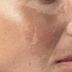
Melasma is a sun-induced hyperpigmentation, usually on the face, that is acquired and appears as light- or dark-brown. In addition to being idiopathic, melasma can also be linked to pregnancy, oral contraceptives, or drugs like diphenylhydantoin. People with darker skin tones tend to have melasma more frequently; women account for 90% of instances. A synonym is chloasma.
a macular hyperpigmentation, usually uniform but sometimes blotchy, that is primarily well defined in the frontal and malar regions of the face.
Clinical diagnosis is done, and post inflammatory hyperpigmentation is included in the differential.
Topical therapies include azelaic acid 20% cream, hydroquinone 3% solution and 4% cream, and a combination of tretinoin 0.05%, hydroquinone 4%, and flucinolone 0.01%.
The pharmacist can combine hydroquinone 4% cream with glycolic acid cream or 0.05% tretinoin cream. Use of monobenzylether or other hydroquinone ethers (monomethyl- or monoethyl-) should be avoided as these ethers might produce a disfiguring patchy leukoderma and result in a permanent loss of melanocytes. Sunscreens can help prevent melasma.
Melasma is a sun-induced hyperpigmentation, usually on the face, that is acquired and appears as light- or dark-brown. In addition to being idiopathic, melasma can also be linked to pregnancy, oral contraceptives, or drugs like diphenylhydantoin. People with darker skin tones tend to have melasma more frequently; women account for 90% of instances. A synonym is chloasma.
a macular hyperpigmentation, usually uniform but sometimes blotchy, that is primarily well defined in the frontal and malar regions of the face.
Clinical diagnosis is done, and post inflammatory hyperpigmentation is included in the differential.
Topical therapies include azelaic acid 20% cream, hydroquinone 3% solution and 4% cream, and a combination of tretinoin 0.05%, hydroquinone 4%, and flucinolone 0.01%.
The pharmacist can combine hydroquinone 4% cream with glycolic acid cream or 0.05% tretinoin cream. Use of monobenzylether or other hydroquinone ethers (monomethyl- or monoethyl-) should be avoided as these ethers might produce a disfiguring patchy leukoderma and result in a permanent loss of melanocytes. Sunscreens can help prevent melasma.
- Published on
Dermatology - Non-genital herpes simplex virus (HSV) infection
Following primary infection, HSV persists in sensory ganglia, and outbreaks recur as
immunity lessens. Recurrences are typically without symptoms or of minimal severity, and frequently do not necessitate any treatment.
Nevertheless, in individuals with weakened immune systems, mucocutaneous lesions can become widespread and long-lasting.
The typical causes contributing to the recurrence of herpes labialis include skin or mucosal irritation, menstruation, fever, common cold, and compromised immunological systems.
Primary infections frequently may not exhibit any noticeable symptoms and may only present with regional lymphadenopathy, as well as systemic symptoms such as fever, headache, malaise, and myalgia. The mouth (characterized by gingival erythema, edema, and pain), anogenital area, and hand/fingers are frequent locations for initial HSV infection. Recurrent infections typically present with a prodrome characterized by tingling, itching, or a burning sensation. However, systemic symptoms are typically not present. Common complications following HSV infection include sensory peripheral nerve illnesses such as eczema herpeticum, erythema multiforme, meningitis, and Bell's palsy.
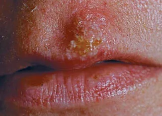
The lesions are red, raised bumps that develop into clusters of fluid-filled blisters and pus-filled bumps that break open easily and create erosions as the top layer of skin sheds. Postinflammatory hyperor hypopigmentation frequently occurs, sometimes resulting in scarring. Oral mucosa lesions typically manifest exclusively during the initial illness.
The diagnosis is made through clinical examination and verified by using a Tzanck smear, viral culture, or antigen detection test.
Seroconversion is used to diagnose first-episode infections. If the patient does not have HSV antibodies in their blood, it can be concluded that they do not have recurring herpes. The differential diagnosis include aphthous stomatitis, hand-foot-and-mouth disease, herpangina, erythema multiforme, and fixed drug eruption.
Advise patients to refrain from engaging in direct touch between their skin surfaces during periods of breakouts.
Systemic oral antivirals are most efficacious in treating first outbreaks: The recommended dosage for Acyclovir is 400 mg taken three times daily, or 200 mg taken five times daily, for a duration of seven to ten days. Famciclovir should be taken at a dosage of 250 mg three times daily for a period of five days. Valacyclovir should be taken at a dosage of 1 g twice daily for a duration of seven to ten days.
Topical antiviral creams and ointments may provide limited efficacy during modest recurrences.
Apply Acyclovir 5% ointment six times daily for a period of seven to 10 days.
Following primary infection, HSV persists in sensory ganglia, and outbreaks recur as
immunity lessens. Recurrences are typically without symptoms or of minimal severity, and frequently do not necessitate any treatment.
Nevertheless, in individuals with weakened immune systems, mucocutaneous lesions can become widespread and long-lasting.
The typical causes contributing to the recurrence of herpes labialis include skin or mucosal irritation, menstruation, fever, common cold, and compromised immunological systems.
Primary infections frequently may not exhibit any noticeable symptoms and may only present with regional lymphadenopathy, as well as systemic symptoms such as fever, headache, malaise, and myalgia. The mouth (characterized by gingival erythema, edema, and pain), anogenital area, and hand/fingers are frequent locations for initial HSV infection. Recurrent infections typically present with a prodrome characterized by tingling, itching, or a burning sensation. However, systemic symptoms are typically not present. Common complications following HSV infection include sensory peripheral nerve illnesses such as eczema herpeticum, erythema multiforme, meningitis, and Bell's palsy.
The lesions are red, raised bumps that develop into clusters of fluid-filled blisters and pus-filled bumps that break open easily and create erosions as the top layer of skin sheds. Postinflammatory hyperor hypopigmentation frequently occurs, sometimes resulting in scarring. Oral mucosa lesions typically manifest exclusively during the initial illness.
The diagnosis is made through clinical examination and verified by using a Tzanck smear, viral culture, or antigen detection test.
Seroconversion is used to diagnose first-episode infections. If the patient does not have HSV antibodies in their blood, it can be concluded that they do not have recurring herpes. The differential diagnosis include aphthous stomatitis, hand-foot-and-mouth disease, herpangina, erythema multiforme, and fixed drug eruption.
Advise patients to refrain from engaging in direct touch between their skin surfaces during periods of breakouts.
Systemic oral antivirals are most efficacious in treating first outbreaks: The recommended dosage for Acyclovir is 400 mg taken three times daily, or 200 mg taken five times daily, for a duration of seven to ten days. Famciclovir should be taken at a dosage of 250 mg three times daily for a period of five days. Valacyclovir should be taken at a dosage of 1 g twice daily for a duration of seven to ten days.
Topical antiviral creams and ointments may provide limited efficacy during modest recurrences.
Apply Acyclovir 5% ointment six times daily for a period of seven to 10 days.
- Published on
Dermatology - Atopic Dermatitis / Atopic Eczema
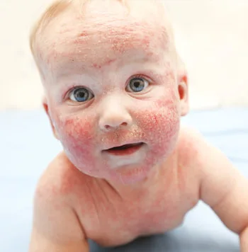
Atopic dermatitis, often known as eczema, typically starts during infancy and reaches its highest occurrence rate of 15-20% in early childhood. Frequently, there exists a familial background of atopy, allergic rhinitis, or asthma.
Triggers encompass aeroallergens, particularly dust mites and pollen; microorganisms; autoallergens; food items, notably eggs, milk, peanuts, soybeans, fish, and wheat; clothes, specifically wool.
Flares can be triggered by stress and are more prevalent during the winter season and after disrobing.
Pruritus is an essential characteristic of eczema, often described as "the itch that rashes." The persistent act of scratching results in a relentless loop of itch → scratch → rash → itch → scratch.
Abnormalities
Erythematous patches, papules, and plaques with ill-defined borders may exhibit scaling and edema. Scratching can lead to the formation of moist and crusty erosions, which may indicate a secondary infection if they are oozing.
Chronic cases might result in the development of lichenification and fissures, which can cause pain. Follicular lichenification can develop, particularly in individuals with deeply pigmented skin. Friction can lead to eyebrow hair loss, darkening of the skin around the eyes, and the formation of a fold below the eyelids known as the Dennie-Morgan sign.
The diagnosis is established through an evaluation of the patient's medical history and a thorough clinical examination. The differential diagnosis comprises seborrheic dermatitis, contact dermatitis, psoriasis, nummular eczema, dermatophytosis, and early mycosis fungoides. Additional uncommon conditions to consider are acrodermatitis enteropathica, glucagonoma syndrome, histidinemia, phenylketonuria, and some immunologic abnormalities.
Instruct patients to refrain from rubbing and scratching, utilize emollients, and take measures to prevent subsequent infection. Administer wet dressings, topical glucocorticoids, and topical antibiotics as necessary, and utilize hydroxyzine to alleviate pruritus. To treat subacute and chronic cases, recommend bathing with oil or oatmeal powder, using unscented emollients, and applying topical anti-inflammatories like glucocorticoids, hydroxyquinoline, or tar. Glucocorticoids exhibit the highest level of effectiveness, while their extended usage may result in negative effects. Tacrolimus and pimecrolimus are highly effective in treating mild exacerbations and subacute cases of atopic dermatitis. Ultraviolet A-Ultraviolet B phototherapy or narrow band UV (311 nm) and PUVA photochemotherapy may also provide positive results.
Atopic dermatitis, often known as eczema, typically starts during infancy and reaches its highest occurrence rate of 15-20% in early childhood. Frequently, there exists a familial background of atopy, allergic rhinitis, or asthma.
Triggers encompass aeroallergens, particularly dust mites and pollen; microorganisms; autoallergens; food items, notably eggs, milk, peanuts, soybeans, fish, and wheat; clothes, specifically wool.
Flares can be triggered by stress and are more prevalent during the winter season and after disrobing.
Pruritus is an essential characteristic of eczema, often described as "the itch that rashes." The persistent act of scratching results in a relentless loop of itch → scratch → rash → itch → scratch.
Abnormalities
Erythematous patches, papules, and plaques with ill-defined borders may exhibit scaling and edema. Scratching can lead to the formation of moist and crusty erosions, which may indicate a secondary infection if they are oozing.
Chronic cases might result in the development of lichenification and fissures, which can cause pain. Follicular lichenification can develop, particularly in individuals with deeply pigmented skin. Friction can lead to eyebrow hair loss, darkening of the skin around the eyes, and the formation of a fold below the eyelids known as the Dennie-Morgan sign.
The diagnosis is established through an evaluation of the patient's medical history and a thorough clinical examination. The differential diagnosis comprises seborrheic dermatitis, contact dermatitis, psoriasis, nummular eczema, dermatophytosis, and early mycosis fungoides. Additional uncommon conditions to consider are acrodermatitis enteropathica, glucagonoma syndrome, histidinemia, phenylketonuria, and some immunologic abnormalities.
Instruct patients to refrain from rubbing and scratching, utilize emollients, and take measures to prevent subsequent infection. Administer wet dressings, topical glucocorticoids, and topical antibiotics as necessary, and utilize hydroxyzine to alleviate pruritus. To treat subacute and chronic cases, recommend bathing with oil or oatmeal powder, using unscented emollients, and applying topical anti-inflammatories like glucocorticoids, hydroxyquinoline, or tar. Glucocorticoids exhibit the highest level of effectiveness, while their extended usage may result in negative effects. Tacrolimus and pimecrolimus are highly effective in treating mild exacerbations and subacute cases of atopic dermatitis. Ultraviolet A-Ultraviolet B phototherapy or narrow band UV (311 nm) and PUVA photochemotherapy may also provide positive results.