- Published on
Dermatology - Trichilemmal Cyst
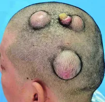
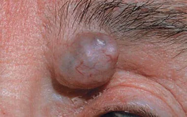
A trichilemmal cyst, which is the second most prevalent type of skin cyst, is commonly observed in middle-aged individuals, particularly women. Familial occurrences of numerous lesions are common.
The majority of trichilemmal cysts, more than 90%, are found on the scalp. The hair on the scalp above the cyst is often unaffected, although it may appear thinner if the cyst is of significant size.
Cysts are characterized by their smooth, hard, dome-shaped appearance, ranging in size from 0.5 to 5 centimeters. They are nodules or tumors that lack a central punctum or attachment to the epidermis. The cyst wall typically exhibits significant thickness, allowing for the complete removal of the cyst in its entirety. The interior of the object consists of keratin, a substance that is highly compact, uniform, and frequently hardened through calcification, featuring cholesterol clefts. In the event of cyst rupture, it can become inflamed and cause intense discomfort.
The diagnosis is determined through clinical examination, and the possible alternatives include keloids, scars, epidermoid cysts, epidermal inclusion cysts, and lipomas.
Tricholemmal cysts are non-cancerous and do not necessitate treatment; however, they can be easily eliminated through surgical excision if desired.
A trichilemmal cyst, which is the second most prevalent type of skin cyst, is commonly observed in middle-aged individuals, particularly women. Familial occurrences of numerous lesions are common.
The majority of trichilemmal cysts, more than 90%, are found on the scalp. The hair on the scalp above the cyst is often unaffected, although it may appear thinner if the cyst is of significant size.
Cysts are characterized by their smooth, hard, dome-shaped appearance, ranging in size from 0.5 to 5 centimeters. They are nodules or tumors that lack a central punctum or attachment to the epidermis. The cyst wall typically exhibits significant thickness, allowing for the complete removal of the cyst in its entirety. The interior of the object consists of keratin, a substance that is highly compact, uniform, and frequently hardened through calcification, featuring cholesterol clefts. In the event of cyst rupture, it can become inflamed and cause intense discomfort.
The diagnosis is determined through clinical examination, and the possible alternatives include keloids, scars, epidermoid cysts, epidermal inclusion cysts, and lipomas.
Tricholemmal cysts are non-cancerous and do not necessitate treatment; however, they can be easily eliminated through surgical excision if desired.
- Published on
Dermatology - Tinea Unguium
Tinea unguium is a fungal infection of the nails that typically does not go away on its own.
The onset often occurs throughout maturity, and the frequency of occurrence seems to rise with age. Transmission occurs by direct contact or contact with contaminated objects, known as fomites. This mode of transmission is most typically observed among family members.
The nails lose their protective and manipulative functions, which can result in pain in the toenail when pressure is applied by shoes. This can also lead to secondary bacterial infections and ulcerations of the underlying nail bed. Complications are more prevalent among individuals with impaired immune systems and those with diabetes. Toenails are more frequently affected than fingernails. Fingernail involvement typically occurs on one side. The typical pattern including fingernails consists of two feet and one hand.
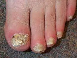
An area of discoloration, typically with clear boundaries, appears on the lower surface of the nail and nail bed. Initially white, it gradually changes to a brown or black color. The nail can get progressively affected over a few weeks or gradually over several months or even years. The nail undergoes increasing infection, resulting in opacity, thickening, cracking, fragility, and elevation due to the accumulation of hyperkeratotic material in the hyponychium. The white streaks, clearly defined and starting at the outer edge of the nail and spreading towards the base, are packed with a combination of keratin debris and air.
The diagnosis is established by confirming the clinical signs through direct microscopic examination of nail clippings using potassium hydroxide preparation, and/or by isolating the pathogenic fungus through culture.
The differential diagnosis comprises psoriatic nails (characterized by "oil drop" staining of the distal nail bed and nail pits, which is observed in psoriasis but not onychomycosis), paronychial psoriasis or eczema, Reiter syndrome and keratoderma blennorrhagicum, onychogryphosis, pincer nails, congenital nail dystrophies, and nail trauma.
Advise the patient to remove damaged nails and administer oral antifungal medication (terbinafine 250 mg per day for 6-16 weeks). The medication can be discontinued once the KOH preparation or culture test shows no signs of infection, even if the nail lesions have not completely healed yet due to the sluggish nail development.
Tinea unguium is a fungal infection of the nails that typically does not go away on its own.
The onset often occurs throughout maturity, and the frequency of occurrence seems to rise with age. Transmission occurs by direct contact or contact with contaminated objects, known as fomites. This mode of transmission is most typically observed among family members.
The nails lose their protective and manipulative functions, which can result in pain in the toenail when pressure is applied by shoes. This can also lead to secondary bacterial infections and ulcerations of the underlying nail bed. Complications are more prevalent among individuals with impaired immune systems and those with diabetes. Toenails are more frequently affected than fingernails. Fingernail involvement typically occurs on one side. The typical pattern including fingernails consists of two feet and one hand.
An area of discoloration, typically with clear boundaries, appears on the lower surface of the nail and nail bed. Initially white, it gradually changes to a brown or black color. The nail can get progressively affected over a few weeks or gradually over several months or even years. The nail undergoes increasing infection, resulting in opacity, thickening, cracking, fragility, and elevation due to the accumulation of hyperkeratotic material in the hyponychium. The white streaks, clearly defined and starting at the outer edge of the nail and spreading towards the base, are packed with a combination of keratin debris and air.
The diagnosis is established by confirming the clinical signs through direct microscopic examination of nail clippings using potassium hydroxide preparation, and/or by isolating the pathogenic fungus through culture.
The differential diagnosis comprises psoriatic nails (characterized by "oil drop" staining of the distal nail bed and nail pits, which is observed in psoriasis but not onychomycosis), paronychial psoriasis or eczema, Reiter syndrome and keratoderma blennorrhagicum, onychogryphosis, pincer nails, congenital nail dystrophies, and nail trauma.
Advise the patient to remove damaged nails and administer oral antifungal medication (terbinafine 250 mg per day for 6-16 weeks). The medication can be discontinued once the KOH preparation or culture test shows no signs of infection, even if the nail lesions have not completely healed yet due to the sluggish nail development.
- Published on
Dermatology - Kawasaki Disease
Kawasaki disease is a sudden and intense sickness characterized by fever in infants and children.
Phase I: Acute Febrile Period: The sudden start of a prolonged period of high fever lasting approximately 12 days, accompanied by symptoms such as diarrhea, arthritis, swollen lymph nodes, and sensitivity to light.
Phase II, known as the Subacute Phase, poses the greatest danger of sudden death and lasts for a further 2-3 weeks. During this phase, individuals experience symptoms such as fever, increased platelet count, skin peeling, joint inflammation, joint pain, and inflammation of the heart.
Phase III, known as the Convalescent Period, occurs 8-10 weeks following the commencement of the illness. During this phase, all indications of the sickness completely disappear and the death rate is minimal. The cessation of disease occurs when the eosinophil sedimentation rate reverts to its normal level.
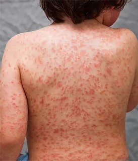
The erythema typically begins on the palms and soles and then spreads to the trunk and extremities, appearing as red patches that can develop into hives-like lesions (most frequently observed), a rash resembling measles (common), or a rash resembling scarlet fever or erythema multiforme (less than 5% of cases). The perineum continues to exhibit persistent erythema in the form of confluent macules progressing to plaque-type lesions, even after other symptoms have resolved. Hand/foot edema is characterized by a pronounced and darkened swelling, accompanied by redness, and affecting the fingers in a spindle-like manner.
The bulbar conjunctival injection commences 48 hours after the start of fever and persists for the duration of the fever.
The lips exhibit a red coloration, are dry, and have cracks, accompanied by widespread redness of the oral cavity and a tongue resembling a "strawberry". Desquamation occurs after a fever, starting at the junction of the nails and the tips of the fingers and toes. Then, layers of the epidermis on the palms and soles of the feet are lost. During the recovery phase, one may observe transverse grooves on the nails.
The diagnosis is established when an individual experiences a fever lasting for five consecutive days, with temperature spikes exceeding 39.4°C, and no other identifiable cause. Additionally, at least four out of five specific criteria must be met. The symptoms include: (1) redness in both eyes; (2) red and cracked lips or throat, or a tongue that looks like a strawberry; (3) redness and swelling of the hands, feet, or all over the body, or peeling skin around the nails; (4) a widespread rash that looks like scarlet fever or red bumps, and eye inflammation; and (5) swollen lymph nodes in the neck measuring at least 1.5 cm. The differential diagnosis comprises various conditions such as adverse cutaneous drug eruptions, juvenile rheumatoid arthritis, infectious mononucleosis, viral exanthems, leptospirosis, Rocky Mountain spotted fever, toxic shock syndrome, staphylococcal scalded-skin syndrome, erythema multiforme, serum sickness, lupus, and reactive arthritis.
Admit the patient to the hospital and closely observe for any cardiovascular complications. Administer intravenous immunoglobulin at a dosage of 2 grams per kilogram in a single infusion lasting 10 hours. Additionally, provide aspirin at a dosage of 100 milligrams per kilogram per day until the fever subsides or until day 14. Afterward, reduce the aspirin dosage to 5 to 10 milligrams per kilogram per day until the eosinophil sedimentation rate returns to normal. Glucocorticoids are linked to an increased incidence of coronary aneurysms and are hence contraindicated.
Kawasaki disease is a sudden and intense sickness characterized by fever in infants and children.
Phase I: Acute Febrile Period: The sudden start of a prolonged period of high fever lasting approximately 12 days, accompanied by symptoms such as diarrhea, arthritis, swollen lymph nodes, and sensitivity to light.
Phase II, known as the Subacute Phase, poses the greatest danger of sudden death and lasts for a further 2-3 weeks. During this phase, individuals experience symptoms such as fever, increased platelet count, skin peeling, joint inflammation, joint pain, and inflammation of the heart.
Phase III, known as the Convalescent Period, occurs 8-10 weeks following the commencement of the illness. During this phase, all indications of the sickness completely disappear and the death rate is minimal. The cessation of disease occurs when the eosinophil sedimentation rate reverts to its normal level.
The erythema typically begins on the palms and soles and then spreads to the trunk and extremities, appearing as red patches that can develop into hives-like lesions (most frequently observed), a rash resembling measles (common), or a rash resembling scarlet fever or erythema multiforme (less than 5% of cases). The perineum continues to exhibit persistent erythema in the form of confluent macules progressing to plaque-type lesions, even after other symptoms have resolved. Hand/foot edema is characterized by a pronounced and darkened swelling, accompanied by redness, and affecting the fingers in a spindle-like manner.
The bulbar conjunctival injection commences 48 hours after the start of fever and persists for the duration of the fever.
The lips exhibit a red coloration, are dry, and have cracks, accompanied by widespread redness of the oral cavity and a tongue resembling a "strawberry". Desquamation occurs after a fever, starting at the junction of the nails and the tips of the fingers and toes. Then, layers of the epidermis on the palms and soles of the feet are lost. During the recovery phase, one may observe transverse grooves on the nails.
The diagnosis is established when an individual experiences a fever lasting for five consecutive days, with temperature spikes exceeding 39.4°C, and no other identifiable cause. Additionally, at least four out of five specific criteria must be met. The symptoms include: (1) redness in both eyes; (2) red and cracked lips or throat, or a tongue that looks like a strawberry; (3) redness and swelling of the hands, feet, or all over the body, or peeling skin around the nails; (4) a widespread rash that looks like scarlet fever or red bumps, and eye inflammation; and (5) swollen lymph nodes in the neck measuring at least 1.5 cm. The differential diagnosis comprises various conditions such as adverse cutaneous drug eruptions, juvenile rheumatoid arthritis, infectious mononucleosis, viral exanthems, leptospirosis, Rocky Mountain spotted fever, toxic shock syndrome, staphylococcal scalded-skin syndrome, erythema multiforme, serum sickness, lupus, and reactive arthritis.
Admit the patient to the hospital and closely observe for any cardiovascular complications. Administer intravenous immunoglobulin at a dosage of 2 grams per kilogram in a single infusion lasting 10 hours. Additionally, provide aspirin at a dosage of 100 milligrams per kilogram per day until the fever subsides or until day 14. Afterward, reduce the aspirin dosage to 5 to 10 milligrams per kilogram per day until the eosinophil sedimentation rate returns to normal. Glucocorticoids are linked to an increased incidence of coronary aneurysms and are hence contraindicated.
- Published on
Dermatology - Erosion
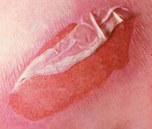
An erosion is a superficial skin lesion that affects only the outer layer (epidermis) and does not extend into the deeper layer (dermis).
The erosion is distinctly delineated, crimson in color, and exuding fluid.
The superficial erosions are either subcorneal or extend into the epidermis.
The papillary body has deep erosions at its base. Erosions are not detectable with palpation.
With the exception of physical abrasions, erosions are always caused by the separation of layers within the skin, either within the outermost layer or just below it, resulting in the formation of vesicles or bullae. Erosions consistently undergo complete healing without leaving any visible scar.
An erosion is a superficial skin lesion that affects only the outer layer (epidermis) and does not extend into the deeper layer (dermis).
The erosion is distinctly delineated, crimson in color, and exuding fluid.
The superficial erosions are either subcorneal or extend into the epidermis.
The papillary body has deep erosions at its base. Erosions are not detectable with palpation.
With the exception of physical abrasions, erosions are always caused by the separation of layers within the skin, either within the outermost layer or just below it, resulting in the formation of vesicles or bullae. Erosions consistently undergo complete healing without leaving any visible scar.
- Published on
Dermatology - Ulcer
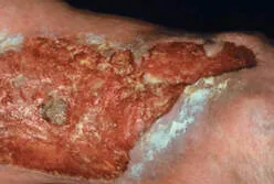
The term "ulcer" is derived from the Latin word for sore. An ulcer is a skin lesion that penetrates into the dermis or subcutis.
Ulcers are invariably a consequence of pathologically modified tissue.
Ulcers can exhibit raised, eroded, indurated, or macerated margins.
Ulcers can exude or exhibit purulent discharge.
The pathologically modified tissue that leads to an ulcer is typically observed at the edge or the foundation of the ulcer, and it aids in identifying the underlying cause of the ulcer. The identification of any accompanying topographic characteristics, such as nodules, excoriations, varicosities, hair distribution, perspiration, and arterial pulses, can aid in determining the underlying etiology. Ulcers invariably undergo scar development during the healing process.
The term "ulcer" is derived from the Latin word for sore. An ulcer is a skin lesion that penetrates into the dermis or subcutis.
Ulcers are invariably a consequence of pathologically modified tissue.
Ulcers can exhibit raised, eroded, indurated, or macerated margins.
Ulcers can exude or exhibit purulent discharge.
The pathologically modified tissue that leads to an ulcer is typically observed at the edge or the foundation of the ulcer, and it aids in identifying the underlying cause of the ulcer. The identification of any accompanying topographic characteristics, such as nodules, excoriations, varicosities, hair distribution, perspiration, and arterial pulses, can aid in determining the underlying etiology. Ulcers invariably undergo scar development during the healing process.
- Published on
Dermatology - Scar
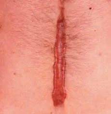
A scar is the fibrous tissue that has grown in place of a previous wound or ulcer.
• Hard, hypertrophic scars are possible.
• Soft, atrophic scars are possible.
• The skin's tissue compartments may diminish or disappear entirely; scars may or may not be noticeable.
A scar is the fibrous tissue that has grown in place of a previous wound or ulcer.
• Hard, hypertrophic scars are possible.
• Soft, atrophic scars are possible.
• The skin's tissue compartments may diminish or disappear entirely; scars may or may not be noticeable.
- Published on
Dermatology - Atrophy
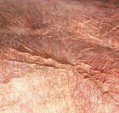
Atrophy is the term used to describe a reduction in size or thickness of one or more layers of the skin.
Abnormalities
Epidermal atrophy is characterized by a reduction in the thickness of the epidermis.
• The outer layer of the skin becomes see-through, exposing the blood vessels in the upper and lower layers of the skin.
• Skin texture is diminished and there are wrinkles resembling cigarette paper. • Dermal atrophy involves the loss of connective tissue in the dermis, resulting in a depressed lesion.
Atrophy is the term used to describe a reduction in size or thickness of one or more layers of the skin.
Abnormalities
Epidermal atrophy is characterized by a reduction in the thickness of the epidermis.
• The outer layer of the skin becomes see-through, exposing the blood vessels in the upper and lower layers of the skin.
• Skin texture is diminished and there are wrinkles resembling cigarette paper. • Dermal atrophy involves the loss of connective tissue in the dermis, resulting in a depressed lesion.
- Published on
Dermatology - Cyst
Cyst
A cyst is an enclosed sac filled with either liquid, solid, or semi-solid substances.
Cysts can be either superficial or deep. Cysts are often round and have a morphology similar to a papule or nodule. When touched, they feel firm and springy.
Cysts are encased by an epithelial layer and frequently possess a fibrous capsule.The color of the cyst might vary depending on its contents, ranging from skin-colored to yellow, red, or blue. An epidermal cyst and a pilar cyst both generate keratinaceous substance.
Cyst
A cyst is an enclosed sac filled with either liquid, solid, or semi-solid substances.
Cysts can be either superficial or deep. Cysts are often round and have a morphology similar to a papule or nodule. When touched, they feel firm and springy.
Cysts are encased by an epithelial layer and frequently possess a fibrous capsule.The color of the cyst might vary depending on its contents, ranging from skin-colored to yellow, red, or blue. An epidermal cyst and a pilar cyst both generate keratinaceous substance.
- Published on
Dermatology -Superficial spreading Melanoma
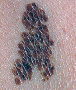
Superficial Spreading melanoma ( SSM)
Light-skinned individuals are more likely to develop SSM melanoma (70%), which typically appears on the upper back. It is a morphologically unique lesion that grows very slowly.
Lesions can happen anywhere, at any age, and are typically solitary. The typical age of onset for SSM is between 30 and 50 years old, and it is slightly more common in women than in persons with darker complexion. Precursor lesions, pale skin, childhood sunburns, and a family history of melanoma are risk factors.
There is a raised, level plaque with distinct uneven boundaries and asymmetry.
The pigment variation of SSM is comparable to, but more pronounced than, the color variability found in the majority of LMM. Lesions are tan, brown, or black. A variety of colors, including red, blue, slate-gray, and dark brown, are displayed, along with areas of tumor regression that are gray or slate-gray. The majority are larger than 5 mm at discovery and develop in pigmented lesions that were previously present (dysplastic nevi). The appearance of nodules and papules indicates a shift to vertical development and dermal invasion.
Dermoscopy and biopsy are used to confirm the clinical diagnosis. When complete excisional biopsy is not possible, incisional or punch biopsy can be carried out; however, total excisional biopsy with small margins is the preferred method. Shave biopsies that don't indicate the extent of invasion should not be used.
Treatment consists of fascia-level excision. Make sure that lesion margins are 1 cm for lesions less than 1 mm in thickness, and only biopsy lymph nodes if they can be felt. Make sure the sentinel lymph nodes are biopsied for lesions thicker than 1 mm. After excision, use skin grafts for repair or direct closure. Only remove lymph nodes if they are clinically palpable and show signs of malignancy, or if there are nodal basins containing concealed tumor cells. If there is a chance of recurrence (positive regional lymph nodes, advanced stage), take adjuvant therapy into consideration.
Superficial Spreading melanoma ( SSM)
Light-skinned individuals are more likely to develop SSM melanoma (70%), which typically appears on the upper back. It is a morphologically unique lesion that grows very slowly.
Lesions can happen anywhere, at any age, and are typically solitary. The typical age of onset for SSM is between 30 and 50 years old, and it is slightly more common in women than in persons with darker complexion. Precursor lesions, pale skin, childhood sunburns, and a family history of melanoma are risk factors.
There is a raised, level plaque with distinct uneven boundaries and asymmetry.
The pigment variation of SSM is comparable to, but more pronounced than, the color variability found in the majority of LMM. Lesions are tan, brown, or black. A variety of colors, including red, blue, slate-gray, and dark brown, are displayed, along with areas of tumor regression that are gray or slate-gray. The majority are larger than 5 mm at discovery and develop in pigmented lesions that were previously present (dysplastic nevi). The appearance of nodules and papules indicates a shift to vertical development and dermal invasion.
Dermoscopy and biopsy are used to confirm the clinical diagnosis. When complete excisional biopsy is not possible, incisional or punch biopsy can be carried out; however, total excisional biopsy with small margins is the preferred method. Shave biopsies that don't indicate the extent of invasion should not be used.
Treatment consists of fascia-level excision. Make sure that lesion margins are 1 cm for lesions less than 1 mm in thickness, and only biopsy lymph nodes if they can be felt. Make sure the sentinel lymph nodes are biopsied for lesions thicker than 1 mm. After excision, use skin grafts for repair or direct closure. Only remove lymph nodes if they are clinically palpable and show signs of malignancy, or if there are nodal basins containing concealed tumor cells. If there is a chance of recurrence (positive regional lymph nodes, advanced stage), take adjuvant therapy into consideration.
- Published on
Dermatology - Wheals
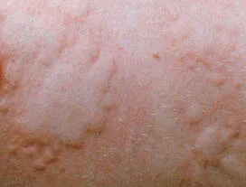
Wheals occur as a result of edema in the papillary layer of the dermis. An eruption characterized by raised, itchy skin lesions is referred to as an urticarial exanthema or urticaria.
A wheal is a transient skin lesion that appears as a pale red papule or plaque with a rounded or flat top, and often disappears within 24-48 hours.
The wheals might have a circular, swirling, or irregular appearance, with fast changing pseudopods that vary in size and shape due to the shifting swelling of the skin's papillary layer.
Wheals can be detected through palpation.
Wheals occur as a result of edema in the papillary layer of the dermis. An eruption characterized by raised, itchy skin lesions is referred to as an urticarial exanthema or urticaria.
A wheal is a transient skin lesion that appears as a pale red papule or plaque with a rounded or flat top, and often disappears within 24-48 hours.
The wheals might have a circular, swirling, or irregular appearance, with fast changing pseudopods that vary in size and shape due to the shifting swelling of the skin's papillary layer.
Wheals can be detected through palpation.